. 2011 Apr 20;6(4):e18932.
doi: 10.1371/journal.pone.0018932.

Lysophosphatidic acid activates peroxisome proliferator activated receptor-γ in CHO cells that over-express glycerol 3-phosphate acyltransferase-1

Affiliations

Lysophosphatidic acid activates peroxisome proliferator activated receptor-γ in CHO cells that over-express glycerol 3-phosphate acyltransferase-1

Cliona M Stapleton et al. PLoS One. .

Abstract

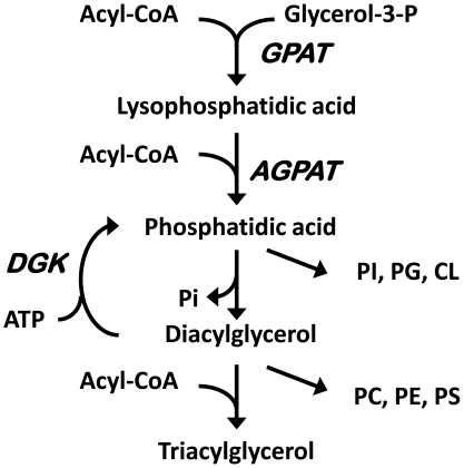
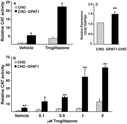
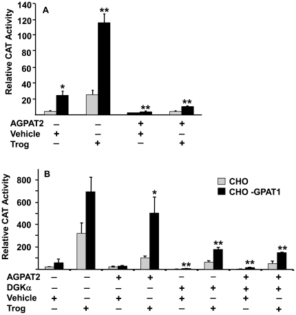
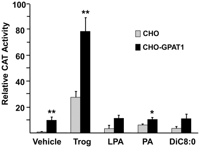
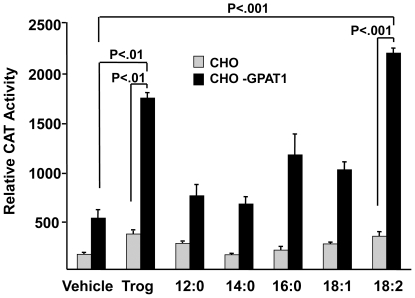
Lysophosphatidic acid (LPA) is an agonist for peroxisome proliferator activated receptor-γ (PPARγ). Although glycerol-3-phosphate acyltransferase-1 (GPAT1) esterifies glycerol-3-phosphate to form LPA, an intermediate in the de novo synthesis of glycerolipids, it has been assumed that LPA synthesized by this route does not have a signaling role. The availability of Chinese Hamster Ovary (CHO) cells that stably overexpress GPAT1, allowed us to analyze PPARγ activation in the presence of LPA produced as an intracellular intermediate. LPA levels in CHO-GPAT1 cells were 6-fold higher than in wild-type CHO cells, and the mRNA abundance of CD36, a PPARγ target, was 2-fold higher. Transactivation assays showed that PPARγ activity was higher in the cells that overexpressed GPAT1. PPARγ activity was enhanced further in CHO-GPAT1 cells treated with the PPARγ ligand troglitazone. Extracellular LPA, phosphatidic acid (PA) or a membrane-permeable diacylglycerol had no effect, showing that PPARγ had been activated by LPA generated intracellularly. Transient transfection of a vector expressing 1-acylglycerol-3-phosphate acyltransferase-2, which converts endogenous LPA to PA, markedly reduced PPARγ activity, as did over-expressing diacylglycerol kinase, which converts DAG to PA, indicating that PA could be a potent inhibitor of PPARγ. These data suggest that LPA synthesized via the glycerol-3-phosphate pathway can activate PPARγ and that intermediates of de novo glycerolipid synthesis regulate gene expression.


Conflict of interest statement

Competing Interests: Two authors, Lisa M. Leesnitzer and Julie B. Stimmel are employees of GlaxoSmithKline. This does not alter our adherence to all the PLoS ONE policies on sharing data and materials.

Figures

Figure 1
Figure 1. Pathway of glycerolipid synthesis.
AGPAT, acyl-glycerol-3-phosphate acyltransferase; CL, cardiolipin; DGK, diacylglycerol kinase; GPAT, glycerol-3-phosphate acyltransferase; PC, phosphatidylcholine; PE, phosphatidylethanolamine; PG, phosphatidylglycerol; PI, phosphatidylinositol; PS, phosphatidylserine.
Figure 2
Figure 2. Overexpression of GPAT1 in CHO cells activated PPARγ.
(A) CHO and CHO-GPAT1 cells were transfected with 0.1 µg of pRLSV40 (internal LUC control) and equal concentrations (0.4 µg) of a PPARγ expression vector, an RXR expression vector, and a PPRE-CAT reporter vector for 24 h, then treated with either 5 µM troglitazone or vehicle (dimethylsulfoxide). (B) CHO and CHO-GPAT1 cells were similarly transfected with the described vectors, then treated with either vehicle or troglitazone (0.1 µM to 5 µM). CAT activity 24 h later was normalized to LUC activity (mean +/- SEM; n = 3) and expressed as relative CAT activity. Results are representative of three and two independent experiments, respectively. (C) CHO and CHO-GPAT1 cells were grown in MEM as described in the methods. RNA was extracted, and CD36 expression was determined by qRTPCR and normalized to GAPDH expression. Data represent the average of four separate experiments. *p<0.05 and **p≤0.01 when comparing within treatment groups.
Figure 3
Figure 3. Adding AGPAT2 and DGKα to CHO-GPAT1 cells decreased PPARγ activity.
(A) CHO and CHO-GPAT1 cells were transfected with 0.1 µg of pRLSV40 (internal LUC control), and equal concentrations (0.4 µg) of expression and reporter vectors, PPARγ RXR, PPRE-CAT, and either empty vector or an AGPAT2 expression vector. (B) Cells were similarly transfected with the vectors described plus either a wild-type (inactive) DGKα expression vector (−) or the constitutively active DGKα Δ196 expression vector (+). Twenty-four hours after transfection, cells were treated with either vehicle or 1 µM troglitazone. CAT activity was measured 24 h later and normalized to LUC activity. The results (mean +/− SEM; n = 3) are expressed as relative CAT activity. Results are representative of three and two independent experiments, respectively. *p<0.05 and **p≤0.01 when comparing within treatment groups.
Figure 4
Figure 4. Treating CHO and CHO-GPAT1 cells with LPA, PA, or DiC8∶0 did not enhance PPARγ activity.
CHO and CHO-GPAT1 cells were transfected with 0.1 μg of pRLSV40 (internal LUC control), and equal concentrations (0.4 μg) of a PPARγ expression vector, an RXR expression vector, and a PPRE-CAT reporter vector. Twenty-four hours later, the transfected cells were treated with vehicle (0.1% BSA), 1 μM troglitazone, 5 µM oleoyl-LPA, 5 µM dioleoyl-PA, or 10 µM DiC8∶0. CAT activity was measured 24 h later and normalized to LUC activity. The results (mean +/− SEM; n = 3) are expressed as relative CAT activity. Results are representative of two independent experiments. *p<0.05 and **p≤0.01 when comparing within treatment groups.
Figure 5
Figure 5. Overexpression of GPAT1 in CHO cells enhanced the effects of fatty acid treatments on PPARγ activity.
CHO and CHO-GPAT1 cells were transfected with 0.1 µg of pRLSV40 (internal LUC control), and equal concentrations (0.4 µg) of a PPARγ expression vector, an RXR expression vector, and a PPRE-CAT reporter vector, as described. Twenty-four hours later, transfected cells were treated with either vehicle (0.1% BSA), 1 µM troglitazone, or 250 µM fatty acid (lauric acid (12∶0), myristic acid (14∶0), palmitic acid (16∶0), oleic acid (18∶1), or linoleic acid (18∶2)). CAT activity was measured 24 h later and normalized to LUC activity. The results (mean +/− SEM; n = 3) are expressed as relative CAT activity and are representative of two independent experiments. Results were analyzed by ANOVA and individual comparisons by Fischers LSD test for multiple comparisons.

Similar articles

Cited by

References

    1. Gervois P, Fruchart J-C, Staels B. Inflammation, dyslipidaemia, diabetes and PPARs: pharmacological interest of dual PPARa and PPARg agonists. Int J Clin Pract. 2004;58:22–29. - PubMed
    1. Berger J, Moller DE. The mechanisms of action of PPARs. Annu Rev Med. 2002;53:409–435. - PubMed
    1. Moolenaar WH, van Meeteren LA, Giepmans BN. The ins and outs of lysophosphatidic acid signaling. Bioessays. 2004;26:870–881. - PubMed
    1. McIntyre TM, Pontsler AV, Silva AR, Hilaire A St, Xu Y, et al. Identification of an intracellular receptor for lysophosphatidic acid (LPA): LPA is a transcellular PPAR agonist. Proc Natl Acad Sci USA. 2003;100:131–136. - PMC - PubMed
    1. Tsukahara T, Tsukahara R, Yasuda S, Makarova N, Valentine WJ, et al. Different residues mediate recognition of 1-O-oleyl-lysophosphatidic acid and rosiglitazone in the ligand binding domain of PPARg. J Biol Chem. 2006;281:3398–3407. - PubMed

Publication types