. 2011 Apr 20;6(4):e18880.
doi: 10.1371/journal.pone.0018880.

Arabidopsis plasmodesmal proteome

Affiliations

Arabidopsis plasmodesmal proteome

Lourdes Fernandez-Calvino et al. PLoS One. .

Abstract

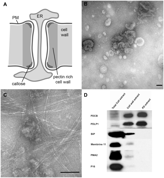
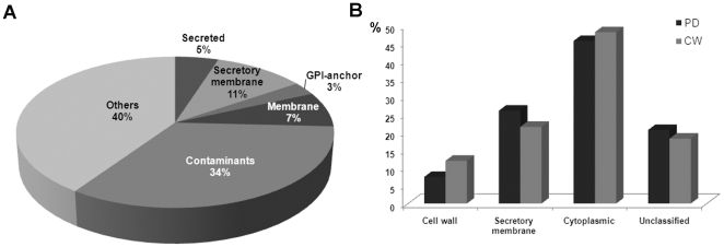
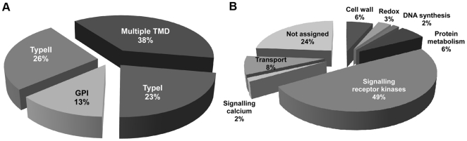
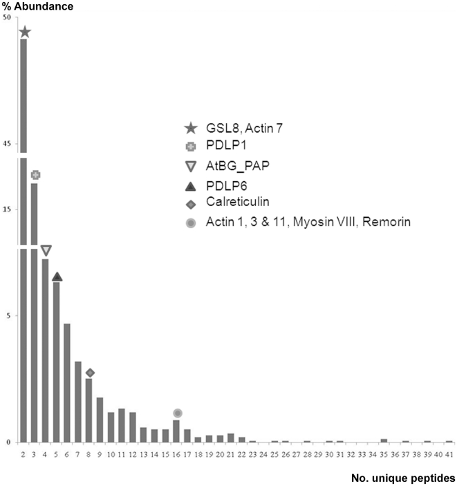
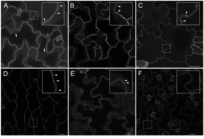
The multicellular nature of plants requires that cells should communicate in order to coordinate essential functions. This is achieved in part by molecular flux through pores in the cell wall, called plasmodesmata. We describe the proteomic analysis of plasmodesmata purified from the walls of Arabidopsis suspension cells. Isolated plasmodesmata were seen as membrane-rich structures largely devoid of immunoreactive markers for the plasma membrane, endoplasmic reticulum and cytoplasmic components. Using nano-liquid chromatography and an Orbitrap ion-trap tandem mass spectrometer, 1341 proteins were identified. We refer to this list as the plasmodesmata- or PD-proteome. Relative to other cell wall proteomes, the PD-proteome is depleted in wall proteins and enriched for membrane proteins, but still has a significant number (35%) of putative cytoplasmic contaminants, probably reflecting the sensitivity of the proteomic detection system. To validate the PD-proteome we searched for known plasmodesmal proteins and used molecular and cell biological techniques to identify novel putative plasmodesmal proteins from a small subset of candidates. The PD-proteome contained known plasmodesmal proteins and some inferred plasmodesmal proteins, based upon sequence or functional homology with examples identified in different plant systems. Many of these had a membrane association reflecting the membranous nature of isolated structures. Exploiting this connection we analysed a sample of the abundant receptor-like class of membrane proteins and a small random selection of other membrane proteins for their ability to target plasmodesmata as fluorescently-tagged fusion proteins. From 15 candidates we identified three receptor-like kinases, a tetraspanin and a protein of unknown function as novel potential plasmodesmal proteins. Together with published work, these data suggest that the membranous elements in plasmodesmata may be rich in receptor-like functions, and they validate the content of the PD-proteome as a valuable resource for the further uncovering of the structure and function of plasmodesmata as key components in cell-to-cell communication in plants.


Conflict of interest statement

Competing Interests: The authors have declared that no competing interests exist.

Figures

Figure 1
Figure 1. Isolation of plasmodesmata.
The basic structure of plasmodesma (PD) is illustrated in Panel A. In addition to the key physical elements of PM, ER, desmotubule in the wall, a speculative arrangement of actin spiralled around the desmotubule is shown. Panel B shows a negatively stained electron micrograph of membranous PD (pellet P2 in M&M) collected after release from the cell wall following cellulase digestion, while Panel C shows contamination of the PD with residual cell wall fibres, observed very occasionally. Scale bars  = 100 nm. Panel D – Immunoblot analysis of fractions harvested during PD isolation procedure. Proteins extracted from whole cells, cell walls (pellet P1 in M&M) and purified PD (pellet P2 in M&M) were analysed using antibodies to the PD marker PDLP1, BiP (ER), Membrine11 (Golgi), PMA2 (PM) and P16 (chloroplast thylakoid envelope). While PDLP was enriched through the isolation procedure, the other proteins diminished and were virtually undetectable in the final PD preparation. Total cell extract: proteins extracted from 6 µl of Arabidopsis cell suspension lysate (corresponding to 0.6 µl of purified cell wall). Cell wall extract: proteins extracted from 75 µl of purified cell walls (pellet P1). PD extract: proteins extracted from 375 µl of purified PD (pellet P2).
Figure 2
Figure 2. Analysis of the PD-proteome with respect to predicted subcellular localization and its comparison with the CW proteome.
(A) The 1341 proteins of the PD-proteome were classified as secreted proteins, integral membrane proteins processed through the secretory pathway and targeted to Golgi, ER, PM and PD (‘secretory membrane’ proteins), GPI-anchor proteins, non-secreted membrane proteins, contaminant proteins and ‘others’, where others are proteins without membrane association and not predicted to be secreted. The contaminant category includes those proteins predicted to be targeted to chloroplasts, mitochondria and vacuoles. Transmembrane helices (using TMHMM [44]), signal peptides (SIGNALP and SIGNALP-HMM [84]), subcellular location (TARGETP [85]) chloroplast transit peptides (CHLOROP [86]), and GPI-anchoring signals (DGPI [87]) were predicted using software as indicated. (B) GO ‘cellular component’ analysis was used to compare the PD-proteome with the previously reported proteomic data for cell walls from Arabidopsis cell cultures (CW). The main cellular component categories; cell wall, secretory membrane, cytoplasmic and unclassified, proteins, were obtained using GO Slim. (The ‘secretory membrane’ class in B is equivalent to the same class in panel A, although it is defined using different software.) ‘Cytoplasmic’ includes plastid, chloroplast, mitochondria, nuclear, ribosome and cytosol proteins. Unclassified category contains other cytoplasmic, other intracellular and unknown cellular categories. Dark gray bars represent PD-proteome and light gray cell wall proteome.
Figure 3
Figure 3. Analysis of membrane proteins targeted to the cell periphery.
Membrane proteins from the PD-proteome were classified into four different categories (A): Type I membrane proteins (Type I), Type II membrane proteins (Type II), GPI- anchor proteins (GPI) and multiple transmembrane domain proteins (Multiple TMD). Panel B shows the predicted functional Mapman categories for the membrane proteins.
Figure 4
Figure 4. Distribution of known PD proteins in the total PD-proteome.
In the hope of identifying potential PD proteins on the basis of the ease of proteomic detection (number of signature peptides), known PD proteins were placed upon a plot of the frequency of identified proteins against detected peptides. No positive correlation was found indicating that PD proteins are very variable in the abundance in PD.
Figure 5
Figure 5. Novel PD proteins identified through their subcellular targeting.
Transgenic expression of fluorescent fusion proteins and their targeting to puncta on the cell wall identified five new PD proteins. Panels A–F show projections of confocal laser scanning microscopy z-series of Arabidopsis leaf epidermal cells for YFP or mRFP fusions to the receptor-like kinases At1g56145(A, YFP), At4g21380(D, YFP), At5g24010 (E, RFP), and At5g59700 (F, YFP), hypothetical protein At3g15480 containing a DUF1218 domain (B, YFP) and a tetraspanin At3g45600 (C, YFP). PD localisation (arrowheads) is evident as punctae of fluorescence in the cell wall for images shown in A–E. For At1g56145 in (A), PD in pitfields at the epidermal-mesophyll boundary are visible (arrows). Panel F illustrates the targeting of a non-PD RLK At5g59700 to the PM. Bars  = 20 µm.

Similar articles

Cited by

References

    1. Lucas WJ, Ham LK, Kim JY. Plasmodesmata - bridging the gap between neighboring plant cells. Trends Cell Biol. 2009;19:495–503. - PubMed
    1. Oparka KJ. Getting the message across: how do plant cells exchange macromolecular complexes? Trends Plant Sci. 2004;9:33–41. - PubMed
    1. Faulkner C, Maule A. Opportunities and successes in the search for plasmodesmal proteins. Protoplasma. 2011;248:27–38. - PubMed
    1. Overall RL, Blackman LM. A model of the macromolecular structure of plasmodesmata. Trends Plant Sci. 1996;1:307–311.
    1. Botha CE, Cross RH. Towards reconciliation of structure with function in plasmodesmata-who is the gatekeeper? Micron. 2000;31:713–721. - PubMed

Publication types