. 2011 Apr 20;6(4):e18877.
doi: 10.1371/journal.pone.0018877.

Towards a synthetic chloroplast

Affiliations

Towards a synthetic chloroplast

Christina M Agapakis et al. PLoS One. .

Abstract

Background: The evolution of eukaryotic cells is widely agreed to have proceeded through a series of endosymbiotic events between larger cells and proteobacteria or cyanobacteria, leading to the formation of mitochondria or chloroplasts, respectively. Engineered endosymbiotic relationships between different species of cells are a valuable tool for synthetic biology, where engineered pathways based on two species could take advantage of the unique abilities of each mutualistic partner.

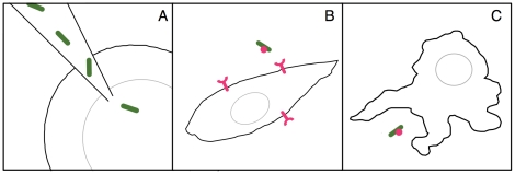
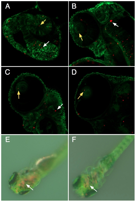
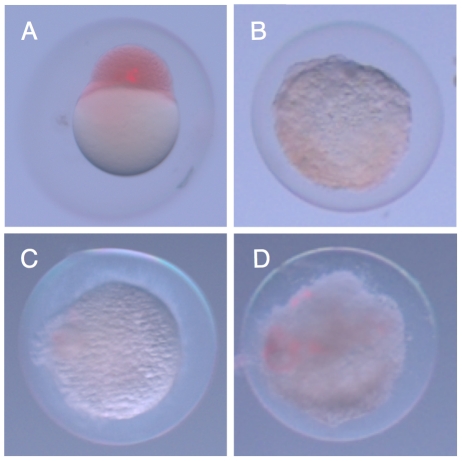
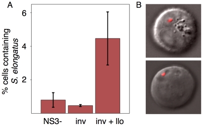
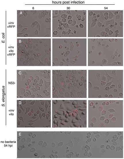
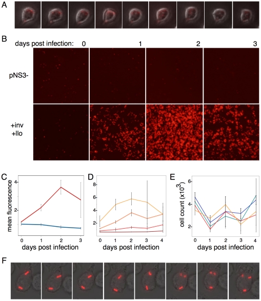
Results: We explored the possibility of using the photosynthetic bacterium Synechococcus elongatus PCC 7942 as a platform for studying evolutionary dynamics and for designing two-species synthetic biological systems. We observed that the cyanobacteria were relatively harmless to eukaryotic host cells compared to Escherichia coli when injected into the embryos of zebrafish, Danio rerio, or taken up by mammalian macrophages. In addition, when engineered with invasin from Yersinia pestis and listeriolysin O from Listeria monocytogenes, S. elongatus was able to invade cultured mammalian cells and divide inside macrophages.

Conclusion: Our results show that it is possible to engineer photosynthetic bacteria to invade the cytoplasm of mammalian cells for further engineering and applications in synthetic biology. Engineered invasive but non-pathogenic or immunogenic photosynthetic bacteria have great potential as synthetic biological devices.


Conflict of interest statement

Competing Interests: The authors have declared that no competing interests exist.

Figures

Figure 1
Figure 1. Three paths to endosymbiosis used in this study.
A.) Direct microinjection of S. elongatus into zebrafish embryos allow exploration of in vivo dynamics of bacteria inside animal cells. B.) Invasion of mammalian cells through heterologous expression of invasin and listeriolysin O. C.) Phagocytosis of bacteria by macrophages. Bacteria subsequently escape from the endosomal compartment through expression of listeriolysin O.
Figure 2
Figure 2. Tracking intracellular S. elongatus through zebrafish development.
Single optical slice confocal microscopy images of the anterior of the zebrafish embryo at A.) Day 1 post injection, B.) Day 2, C.) Day 3, D.) Day 4, and dissecting microscope images of embryos E.) Day 8, F.) Day 12 post injection. Zebrafish cell membranes are outlined in green, with red autofluorescent bacteria visible in cells throughout the embryo, including the eye (yellow arrows) and brain (white arrows). Red autofluorescence gradually decreased over the course of experimental observations, but remained visible in the brain of the young zebrafish even after 12 days.
Figure 3
Figure 3. Zebrafish embryos are immediately killed by E. coli.
A.) Zebrafish embryo two hours after injection of S. elongatus. Cells appear red due to phenol red present in the injection buffer. B.) Injection of E. coli led to drastic morphological changes in the embryo after two hours, and this change was observed with E. coli cells that were C.) UV killed, or D.) ΔmsbB mutants.
Figure 4
Figure 4. Invasion of CHO cells.
A.) S. elongatus engineered with invasin and listeriolysin are able to invade CHO cells at a higher efficiency than S. elongatus harboring the empty vector or invasin alone. Cells positive for red fluorescence were sorted by FACS and B.) observed under confocal microscopy, showing intracellular localization of at least one bacterial cell per CHO cell in the majority of the cells observed.
Figure 5
Figure 5. E. coli and S. elongatus lead to differential effects when phagocytosed by macrophages.
Large scale granulation is observed when macrophages take up E. coli that is A.) not expressing llo and to an even greater extent with B.) E. coli expressing llo off of the inducible lac promoter of the pNS3 vector. In contrast, macrophages displayed similar morphology two days after infection with C.) empty vector S. elongatus, D.) S. elongatus expressing inv and llo, and E.) macrophages untreated with bacteria but maintained at 30°C in bright light for two days.
Figure 6
Figure 6. S. elongatus can grow inside the macrophage cytoplasm.
A.) Time lapse microscopy of macrophages infected with +inv+llo S. elongatus kept in the dark shows the gradual decrease in red autofluorescence over the course of 12 hours. In contrast, when kept in the light, B.) empty vector S. elongatus autofluorescence is observed to gradually decrease over the course of several days (top row), while a significant increase in red S. elongatus autofluorescence was observed in macrophages infected with inv llo S. elongatus for two days post-infection (bottom row). This fluorescence was observed to decrease after the third day of infection. C.) This change in fluorescence over time can be quantified as a change in background subtracted mean fluorescence in ImageJ and averaged over triplicate experiments. Empty vector (blue line) and +inv+llo S. elongatus (red line) show marked differences in growth when infected at similar densities of 1–2 bacterial cells per macrophage. D.) +inv+llo S. elongatus displayed infection density dependent growth rates in macrophages. Each line shows change in mean fluorescence in cells infected at a single starting density, ranging in multiples of two from fewer than one cell per macrophage to approximately 4 bacteria per macrophage. E.) Macrophage cell counts were variable across replicates and over the course of the experiment but displayed no significant difference between macrophages infected with empty vector S. elongatus at low (green line) or high density (blue line), or +inv+llo S. elongatus at low (red line) or high (yellow line) density. F.) When infected at low density of fewer than one bacteria per macrophage, S. elongatus division was observed during 18 hour time-lapse fluorescent microscopy in approximately 1% of macrophages observed, in particular those cells that contained more than one bacterial cell due to stochastic fluctuations.

References

    1. Kleine T, Maier UG, Leister D. DNA Transfer from Organelles to the Nucleus: The Idiosyncratic Genetics of Endosymbiosis. Annu Rev Plant Biol. 2009;60:115–138. - PubMed
    1. Pelz O, Tesar M, Wittich RM, Moore ER, Timmis KN, et al. Towards elucidation of microbial community metabolic pathways: unravelling the network of carbon sharing in a pollutant-degrading bacterial consortium by immunocapture and isotopic ratio mass spectrometry. Environ Microbiol. 1999;1:167–174. - PubMed
    1. Gray M, Burger G, Lang B. Mitochondrial evolution. Science. 1999;283:1476. - PubMed
    1. Howe CJ, Barbrook AC, Nisbet RER, Lockhart PJ, Larkum AWD. The origin of plastids. Philos Trans R Soc Lond, B, Biol Sci. 2008;363:2675–2685. - PMC - PubMed
    1. Douglas A. Nutritional interactions in insect-microbial symbioses: aphids and their symbiotic bacteria Buchnera. Annual Review of Entomology. 1998;43:17–37. - PubMed

Publication types