. 2011 Apr 14;6(4):e18810.
doi: 10.1371/journal.pone.0018810.

Response of estrogen receptor-positive breast cancer tumorspheres to antiestrogen treatments

Affiliations

Response of estrogen receptor-positive breast cancer tumorspheres to antiestrogen treatments

Ada Ao et al. PLoS One. .

Abstract

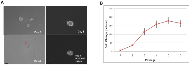
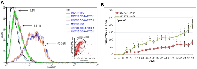
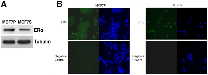
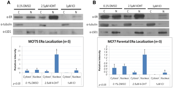
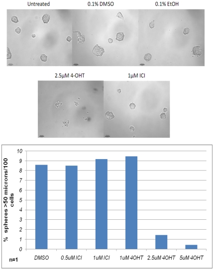
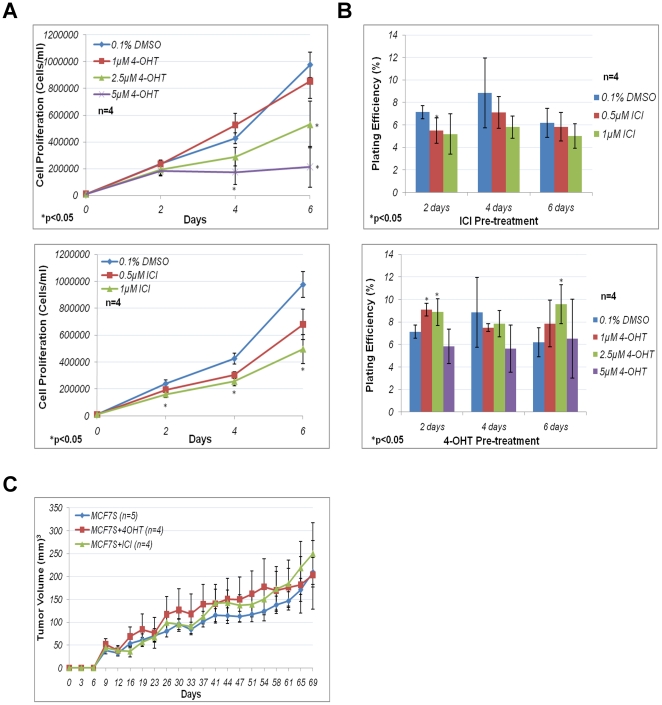
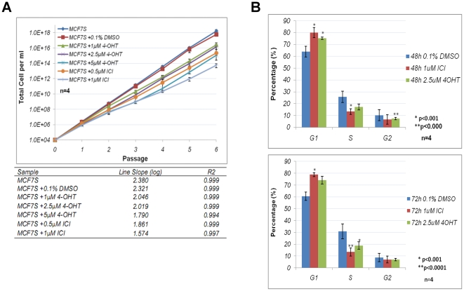
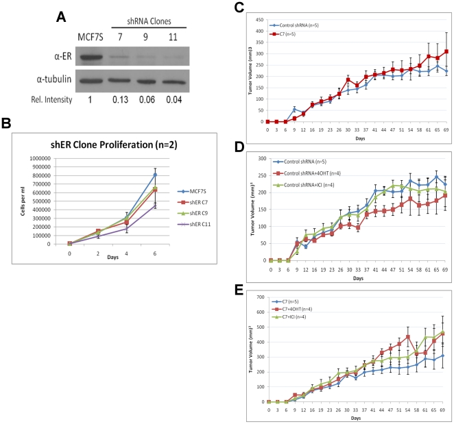
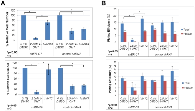
Estrogen signaling plays a critical role in the pathogenesis of breast cancer. Because the majority of breast carcinomas express the estrogen receptor ERα, endocrine therapy that impedes estrogen-ER signaling reduces breast cancer mortality and has become a mainstay of breast cancer treatment. However, patients remain at continued risk of relapse for many years after endocrine treatment. It has been proposed that cancer recurrence may be attributed to cancer stem cells (CSCs)/tumor-initiating cells (TICs). Previous studies in breast cancer have shown that such cells can be enriched and propagated in vitro by culturing the cells in suspension as mammospheres/tumorspheres. Here we established tumorspheres from ERα-positive human breast cancer cell line MCF7 and investigated their response to antiestrogens Tamoxifen and Fulvestrant. The tumorsphere cells express lower levels of ERα and are more tumorigenic in xenograft assays than the parental cells. Both 4-hydroxytamoxifen (4-OHT) and Fulvestrant attenuate tumorsphere cell proliferation, but only 4-OHT at high concentrations interferes with sphere formation. However, treated tumorsphere cells retain the self-renewal capacity. Upon withdrawal of antiestrogens, the treated cells resume tumorsphere formation and their tumorigenic potential remains undamaged. Depletion of ERα shows that ERα is dispensable for tumorsphere formation and xenograft tumor growth in mice. Surprisingly, ERα-depleted tumorspheres display heightened sensitivity to 4-OHT and their sphere-forming capacity is diminished after the drug is removed. These results imply that 4-OHT may inhibit cellular targets besides ERα that are essential for tumorsphere growth, and provide a potential strategy to sensitize tumorspheres to endocrine treatment.


Conflict of interest statement

Competing Interests: The authors have declared that no competing interests exist.

Figures

Figure 1
Figure 1. Establishment of tumorspheres from MCF7.
A. Phase contrast images of tumorspheres derived from MCF7 cells 2, 4, and 8 days after initial seeding. Red arrows (bottom left) indicate microspikes, which are presumed to be microfilaments that spheroid cells use to sense nutrients in the environment. Hoechst nuclei staining (bottom right) shows a multicellular tumorsphere. Magnification at 100x. Scale bar  = 100 microns. B. Growth kinetics of MCF7S. Cells were seeded at 10,000 per ml and allowed to grow for seven days. The cells were then dissociated, counted, and passaged on the seventh day. This was repeated for 6 weeks. The experiment was performed twice with technical triplicates each time. Fold change is shown as final cell density/initial cell density.
Figure 2
Figure 2. Increased tumorigenecity of MCF7S cells.
A. Histogram of CD44-FITC and Iso-FITC staining for MCF7P and MCF7S. Duplicates are shown. Percentages of CD44 positive staining (from 30,000 cells) are indicated. Representative scatter plot and gating of FACS sorted cells is shown as inset. B. In vivo tumorigenic assay for MCF7 parental and MCF7S tumorsphere cells. Five mice were used for each group.
Figure 3
Figure 3. Estrogen receptor status in MCF7S.
A. Immunoblotting of ERα protein in MCF7P and MCF7S cells. Tubulin served as loading control. B. Indirect immunofluorescence for ERα protein (green) in MCF7P and MCF7S. Cells were fixed by 3.7% formaldehyde. HOECHST 33342 (blue) was used to indicate nuclear region. A negative control was performed without primary anti-ERα antibody. Magnification at 40x.
Figure 4
Figure 4. Effects of antiestrogens on ERα abundance and subcellular localization.
Immunoblot of ERα protein in cytoplasmic and nuclear fractions from MCF7S (A) or MCF7 parental cells (B) treated for 48 hours with 4-OHT or ICI. Densitometry quantification of three independent experiments is shown below. Tubulin was used as a loading control for cytoplasmic (C) and LSD1 for nuclear (N) fraction. Statistically analysis was performed using paired Student's t-test.
Figure 5
Figure 5. Effects of antiestrogens on tumorsphere formation.
(Top) Phase-contrast images of MCF7S cells in the presence of antiestrogens or vehicle controls for 7 days. (Bottom) Quantification of MCF7S spheres with >50 micron diameter. Magnification at 100×. Scale bar  = 100 microns.
Figure 6
Figure 6. MCF7S antiestrogen response in vitro.
A. Cell proliferation of MCF7S in the presence of antiestrogens. Cell proliferation of MCF7S treated with 4-OHT (top) or ICI (bottom) from four independent experiments. Error bars represent standard error of the mean (S.E.M.). B. Sphere formation of MCF7S after antiestrogen challenge with 4-OHT or ICI. (Top) Sphere formation frequency of 4-OHT-(top) or ICI-(bottom) treated MCF7S from four independent experiments. Plating efficiency was calculated as number of sphere >50 microns in diameter/total number of cells seeded ×100%. Error bars represent S.E.M. C. In vivo tumorigenic assay for MCF7S cells following antiestrogen challenge. MCF7S were either untreated, treated with 2.5 µM 4-OHT or 1 µM ICI for 4 days then recovered for 6 days in culture without antiestrogens, prior to injections. At least 4 mice were injected for each condition.
Figure 7
Figure 7. Effects of antiestrogens on long-term growth and cell cycle of MCF7S.
A. Long-term expansion of MCF7S in the presence of antiestrogens. The lines are expressed on a semilog graph (top) and slope of each line was calculated as log of averaged expansion (bottom). Data were derived from four independent experiments. Error bars: S.E.M. B. Propidium iodide (PI) cell cycle analysis of 48 (top) and 72 (bottom) hours antiestrogen treated MCF7S. Data were derived from four independent experiments and analyzed using ModFit LT software. Error bars: S.E.M.
Figure 8
Figure 8. ERα is disposable in MCF7S.
A. Immunoblot analysis of ERα in three shERα MCF7S clones. Relative densitometry intensity is shown. B. Cell proliferation of three shERα clones compared to bulk MCF7S culture. Error bars represent standard deviation from two experiments. C. In vivo tumorigenic assay comparing shRNA control and shERα knockdown MCF7S. Five mice were injected for each condition. D and E. In vivo tumorigenic assay comparing antiestrogen treated (4 days) shRNA control or shERα MCF7S cells. At least 4 mice were injected for each condition.
Figure 9
Figure 9. Antiestrogen response of ERα knockdown MCF7S cells.
A. In vitro antiestrogen response of ERα knockdown in MCF7S cells. % Relative Cell Number represents viable cell number in antiestrogen-treated samples relative to vehicle-treated control. Data for two individual clones were generated from three independent experiments. Error bars represent S.E.M. B. Sphere formation frequency of antiestrogen treated ERα knockdown MCF7S cells. Three independent experiments were performed for two individual clones. Error bars: S.E.M.

References

    1. McDonnell DP, Norris JD. Connections and Regulation of the Human Estrogen Receptor. Science. 2002;296:1642–1964. - PubMed
    1. Anderson WF, Chatterjee N, Ershler WB, Brawley OW. Estrogen receptor breast cancer phenotypes in the Surveillance, Epidemiology, and End Results database. Breast Cancer Res Treat. 2002;76:27–36. - PubMed
    1. Ariazi EA, Ariazi JL, Cordera F, Jordan VC. Estrogen receptors as therapeutic targets in breast cancer. Curr Top Med Chem. 2006;6:181–202. - PubMed
    1. Cleator SJ, Ahamed E, Coombes RC, Palmieri C. A 2009 update on the treatment of patients with hormone receptor-positive breast cancer. Clin Breast Cancer. 2009;9(Suppl):1S6–S17. - PubMed
    1. Sengupta S, Jordan VC. Selective estrogen modulators as an anticancer tool: mechanisms of efficiency and resistance. Adv Exp Med Biol. 2008;630:206–219. - PubMed

Publication types