. 2011 Apr 12;6(4):e18503.
doi: 10.1371/journal.pone.0018503.

Regeneration of cryoinjury induced necrotic heart lesions in zebrafish is associated with epicardial activation and cardiomyocyte proliferation

Affiliations

Regeneration of cryoinjury induced necrotic heart lesions in zebrafish is associated with epicardial activation and cardiomyocyte proliferation

Kristin Schnabel et al. PLoS One. .

Abstract

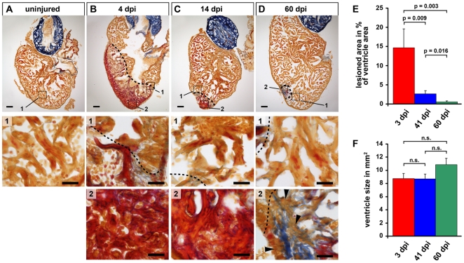
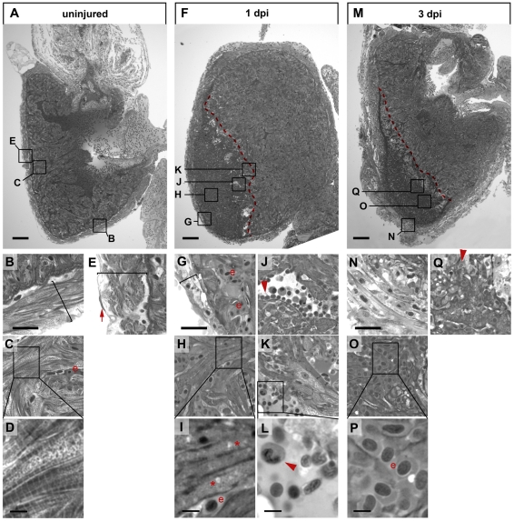
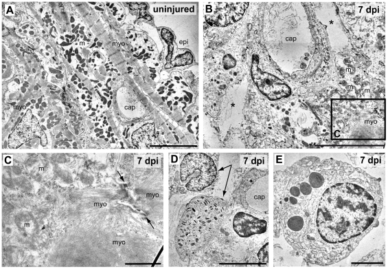
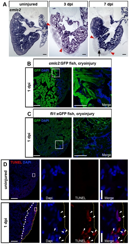
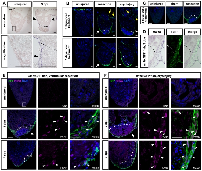
In mammals, myocardial cell death due to infarction results in scar formation and little regenerative response. In contrast, zebrafish have a high capacity to regenerate the heart after surgical resection of myocardial tissue. However, whether zebrafish can also regenerate lesions caused by cell death has not been tested. Here, we present a simple method for induction of necrotic lesions in the adult zebrafish heart based on cryoinjury. Despite widespread tissue death and loss of cardiomyocytes caused by these lesions, zebrafish display a robust regenerative response, which results in substantial clearing of the necrotic tissue and little scar formation. The cellular mechanisms underlying regeneration appear to be similar to those activated in response to ventricular resection. In particular, the epicardium activates a developmental gene program, proliferates and covers the lesion. Concomitantly, mature uninjured cardiomyocytes become proliferative and invade the lesion. Our injury model will be a useful tool to study the molecular mechanisms of natural heart regeneration in response to necrotic cell death.


Conflict of interest statement

Competing Interests: The authors have declared that no competing interests exist.

Figures

Figure 1
Figure 1. The zebrafish heart regenerates after cryoinjury induced tissue damage.
(A–D) Sections of uninjured control hearts (A) and hearts at 4 (B), 14 (C) and 60 (D) days after cryolesion (dpi  =  days post injury) stained with Acid Fuchsin Orange G, which labels cardiomyocytes in orange, fibrin in red and collagen in blue. Magnification #1 of uninjured heart shows healthy myocardium. Magnifications #1 of injured hearts show cardiomyocytes at the lesion edges and parts of wound tissue. Close up views of the lesioned area are shown in magnifications #2. Note that the lesioned area, showing fibrin accumulation at 4 dpi and 14 dpi and minor deposits of collagen at 60 dpi is decreasing in size. Cardiomyocytes are missing at 4 and 14 dpi and at 60 dpi remaining collagen rich tissue is interspersed with cardiomyocytes (arrowheads). Wound edges are indicated by a black dashed line. Scale bars: 100 µm in overviews, 25 µm in close ups. (E) Quantification of the lesioned area normalized to the size of the ventricle. Measurements were performed on all serial sections for each heart. Error bars  =  s.e.m., significance tested by Student's t-test (41 dpi and 60 dpi) and Mann-Whitney rank sum test (others). n = 5 hearts 3 dpi (73 sections), 6 hearts 41dpi (89 sections), 7 hearts 60 dpi (118 sections). (F) Quantification of the ventricular area of the hearts analysed in E. Measurements were done on all sections for each heart. Significance tested by Student's t-test (41 dpi and 60 dpi) and Mann-Whitney rank sum test (others).
Figure 2
Figure 2. Cryoinjury causes cardiomyocyte death, inflammatory response and massive changes in tissue morphology.
Semi-thin histological sections of an uninjured heart (A–E) and cryolesioned hearts at 1 dpi (F–L) and 3 dpi (M–Q) stained with Tuloidin blue are shown. Wound edges are indicated with red dashed lines (F, M). Note striated cardiomyocytes in the compact external (B) and in the trabeculated internal (C, D) myocardium in the uninjured heart and the single layer of epicardial epithelium covering the myocardium (red arrow in E). (F–L) At 1 dpi, the thickness of the external myocardial layer is reduced (compare brackets in B and G) in the lesioned area, and most cardiomyocytes have lost striations suggesting myofibril disassembly (G, H, I). Also note appearance of large vacuolar structures (asterisk in I) indicative of cell death. Leukocytes, largely heterophil granulocytes, delineate the wound edges (J, K, L; see red arrowheads). (M–Q) At 3 dpi the external ventricular layer displays a loosened morphology but is devoid of cardiomyocytes (N). At 3 dpi the lesioned area is largely filled with erythrocytes (“e” in O, P). Cellular debris in the wound area is often associated with granulocytes (red arrowhead in Q). For each time point n = 3 hearts. Scale bars in A, F and M are 100 µm. Scale bars in B, G and N are 25 µm. Scale bars in D, I, L and P are 5 µm.
Figure 3
Figure 3. Ultrastructural analysis of cryolesioned myocardium reveals cardiomyocyte cell death and an inflammatory response.
Electron micrographs of uninjured (A) and cryolesioned (7 dpi, B–E) zebrafish heart tissue. A normal organization of ventricular periphery with epicardal epithelium (epi) and subepicardial cardiomyocytes displaying well-organized striated myofilaments (myo) with Z lines and groups of electron dense mitochondria (m). A capillary (cap) is visible as well. (B) At 7 dpi, the lesioned area displays cellular debris and large tissue gaps (*) around a small capillary. In the lower right corner: cardiomyocyte with damaged mitochondria (m) and disorganised myofilaments (myo). (C) Magnification of the boxed area in B. A cryoinjured cardiomyocyte with disorganised mitochondria (m), myofilaments, and a loosened/ill defined intercalated disc (arrows) is shown. (D, E) Granulocytes in the wound area. (D) Heterophil granulocytes (arrows), the upper one with a peripheral, nonsegmented nucleus, the lower one with characteristic, cigar-shaped cytoplasmic granules. (E) Eosinophil granulocyte. Scale bars: 5 µm in A, B, and D, 1 µm in C, and 2 µm in E.
Figure 4
Figure 4. Cryolesion results in loss of cardiomyocyte and endocardium-specific marker gene expression and in induction of apoptosis.
(A) In situ hybridization shows loss of cmlc2 (myl7) expression in the lesioned area at 3 and 7 dpi. The lesioned area is indicated by red arrowheads. Note the protrusions of cmlc2 positive areas (black arrow) extending into the lesion at 7 dpi. Scale bars are 100 µm. n = 3 hearts (9 sections each) for each uninjured, 3 dpi and 7 dpi. (B) Loss of cmlc2 expression is already evident in cryoinjured hearts at 1 dpi of cmlc2:GFP transgenic fish stained for GFP by immunofluorescence. Nuclei are stained with Dapi. Scale bar in overview is 100 µm, 25 µm in close-up. (C) Endocardial GFP expression is lost in cryolesions of fli1:eGFP transgenic fish at 1 dpi. (D) TUNEL assay on hearts at 1 dpi detects an increased number of apoptotic cells (white arrowheads) compared to uninjured hearts. Wound area is indicated by white dashed line and the periphery of the heart is marked by yellow dashed lines. Nuclei are stained with Dapi. Scale bar in overview is 100 µm. Scale bar in close up is 10 µm. n = 3 hearts each uninjured and 1 dpi.
Figure 5
Figure 5. Organ-wide activation of embryonic gene expression and cell proliferation in the epicardium in response to cryoinjury and ventricular resection.
(A) In situ hybridization for tbx18 shows an organ-wide expression (arrowheads) in the epicardium 3 days after cryoinjury compared to undetectable expression in uninjured hearts. Wound edges are indicated by black dashed lines. Scale bars are 100 µm. n = 3 heart control and 3 dpi. (B) Organ-wide GFP induction in the transgenic wt1b:GFP line shows activated epicardium after ventricular resection and after cryoinjury at 3 and 7 days after treatment. Induction of the transgene is visible in the thickened epicardium near the wound (white arrows) and in the epicardium lining the bulbus arteriosus and atrium as well (yellow arrows). Uninjured hearts show no GFP expression. Nuclei were stained with Dapi. Wound edges are indicated by white dashed lines. Scale bars are 100 µm. n = 5 hearts at all time points. (C) Organ-wide GFP induction in the transgenic wt1b:GFP line is only observed after ventricular resection but not in uninjured or sham amputated control hearts. Nuclei were stained with Dapi. Scale bars are 100 µm. (D) GFP expression in injured wt1b:GFP transgenic hearts is specific to the epicardium. Coexpression of tbx18 transcript detected via in situ hybridization and GFP detected via antibody staining at 3 days post amputation is shown. Scale bar is 100 µm. n = 3 hearts. Dashed line indicates wound edge. (E–F) Activation of epicardial proliferation in response to ventricular resection (E) and cryoinjury (F). Uninjured and amputated hearts at 3 dpa and 7 dpa (E) or cryoinjured hearts at 3 dpi and 7 dpi (F) of wt1b:GFP fish were stained for GFP and PCNA expression. Nuclei are stained with Dapi. Proliferating epicardial cells are indicated with white arrowheads. Note partial coverage of the wound with GFP-positive epicardium in E (arrow) and full coverage in F (arrow). Scale bars are 100 µm in the overview and 25 µm in the close ups. Wound plane is indicated by white dashed lines. n  = 3 hearts (9 sections) for all conditions.
Figure 6
Figure 6. Activation of cardiomyocyte proliferation in response to cryoinjury.
(A) Mature cardiomyocytes located close to the lesion in the uninjured myocardium proliferate at 3 and 7 dpi. Uninjured and cryolesioned hearts (3 dpi and 7 dpi) of cmlc2:GFP transgenic fish were stained for GFP and PCNA. Nuclei are stained with Dapi. Proliferating cardiomyocytes are indicated by white arrowheads. Proliferating endocardial cells are indicated by yellow arrowheads (3 dpi). Note individual GFP+ cardiomyocytes inside the lesioned area at 7 dpi (red arrowheads). Scale bars are 100 µm in the overview and 25 µm in the close ups. n = 3 hearts (9 sections) at all conditions. (B) Quantification of the percentage of PCNA+ cardiomyocytes in uninjured versus cryolesioned hearts at 3 dpi. CMs were quantified in the entire ventricle. Error bars  =  s.e.m. n = 3 hearts (7 sections for 3 dpi, 5260 CMs counted; 9 sections for uninjured control, 7475 CMs counted), Student's t-test was employed for statistical analysis, with p<0.05. (C) Endocardial cells located adjacent to the lesion proliferate at 3 dpi. Cryolesioned hearts (3 dpi) of fli1:eGFP transgenic fish were stained for GFP and PCNA. Nuclei are stained with Dapi. Proliferative endocardial cells are indicated by yellow arrowheads. Wound edge is indicated by a white dashed line. Scale bars are 25 µm.

References

    1. Stoick-Cooper CL, Moon RT, Weidinger G. Advances in signaling in vertebrate regeneration as a prelude to regenerative medicine. Genes & Development. 2007;21:1292–1315. - PubMed
    1. Brockes JP, Kumar A. Comparative aspects of animal regeneration. Annu Rev Cell Dev Biol. 2008;24:525–549. - PubMed
    1. Tanaka EM. Cell differentiation and cell fate during urodele tail and limb regeneration. Curr Opin Genet Dev. 2003;13:497–501. - PubMed
    1. Poss KD, Wilson LG, Keating MT. Heart regeneration in zebrafish. Science. 2002;298:2188–2190. - PubMed
    1. Laube F, Heister M, Scholz C, Borchardt T, Braun T. Re-programming of newt cardiomyocytes is induced by tissue regeneration. J Cell Sci. 2006;119:4719–4729. - PubMed

Publication types

LinkOut - more resources