

Ectopic study of tissue-engineered bone complex with enamel matrix proteins, bone marrow stromal cells in porous calcium phosphate cement scaffolds, in nude mice
- PMID: 21535268
- PMCID: PMC6496638
- DOI: 10.1111/j.1365-2184.2011.00750.x
Ectopic study of tissue-engineered bone complex with enamel matrix proteins, bone marrow stromal cells in porous calcium phosphate cement scaffolds, in nude mice
Abstract
Objective: This study aimed to investigate the potential of enamel matrix proteins (EMPs) on promoting osteogenic differentiation of porcine bone marrow stromal cells (pBMSCs), as well as new bone formation capabilities, in a tissue-engineered bone complex scaffold of EMPs, pBMSCs and porous calcium phosphate cement (CPC).
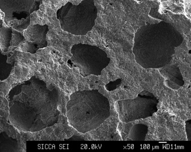
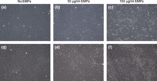
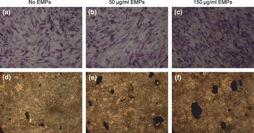
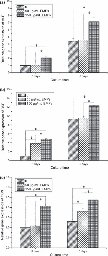
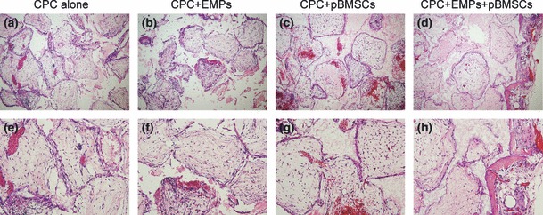
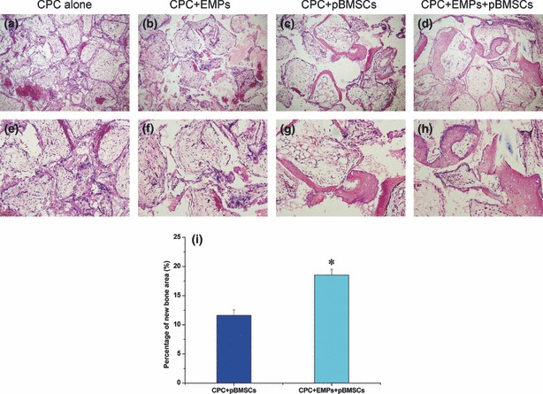
Materials and methods: Effects of EMPs on pBMSCs in vitro was first determined by alkaline phosphatase (ALP) activity, von Kossa staining assay and mRNA expression of ALP, bone sialoprotein (BSP) and osteocalcin (OCN) genes. Next, an ectopic new bone formation test was performed in a nude mouse model with four groups: CPC scaffold alone; CPC scaffold + EMPs; CPC scaffold + pBMSCs; and CPC scaffold + EMPs + pBMSCs, for 2 or 4 weeks.
Results: ALP activity, von Kossa assay and mRNA expressions of ALP, BSP and OCN genes were all significantly higher with 150 μg/ml EMP treatment in vitro. In nude mice, new bone formation was detected only in the CPC scaffold + EMPs + pBMSCs group at 2 weeks. At 4 weeks, in the tissue-engineered construct there was significantly higher bone formation ability than other groups.
Conclusions: EMPs promoted osteogenic differentiation of pBMSCs, and the tissue-engineered complex of EMPs, pBMSCs and CPC scaffold may be a valuable alternative to be used in periodontal bone tissue engineering and regeneration.
© 2011 Blackwell Publishing Ltd.
Figures
Similar articles
-
Minipig-BMSCs Combined with a Self-Setting Calcium Phosphate Paste for Bone Tissue Engineering.Mol Biotechnol. 2016 Nov;58(11):748-756. doi: 10.1007/s12033-016-9974-6. Mol Biotechnol. 2016. PMID: 27683256
-
Construction of vascularized tissue-engineered bone with a double-cell sheet complex.Acta Biomater. 2018 Sep 1;77:212-227. doi: 10.1016/j.actbio.2018.07.024. Epub 2018 Jul 12. Acta Biomater. 2018. PMID: 30017924
-
[A novel tissue-engineered bone constructed by using human adipose-derived stem cells and biomimetic calcium phosphate scaffold coprecipitated with bone morphogenetic protein-2].Beijing Da Xue Xue Bao Yi Xue Ban. 2017 Feb 18;49(1):6-15. Beijing Da Xue Xue Bao Yi Xue Ban. 2017. PMID: 28202997 Chinese.
-
Maxillary sinus floor elevation using a tissue-engineered bone with calcium-magnesium phosphate cement and bone marrow stromal cells in rabbits.Tissue Eng Part A. 2012 Apr;18(7-8):870-81. doi: 10.1089/ten.TEA.2011.0379. Epub 2011 Dec 22. Tissue Eng Part A. 2012. PMID: 22066969
-
Calcium Phosphates: A Key to Next-Generation In Vitro Bone Modeling.Adv Healthc Mater. 2024 Nov;13(29):e2401307. doi: 10.1002/adhm.202401307. Epub 2024 Aug 23. Adv Healthc Mater. 2024. PMID: 39175382 Free PMC article. Review.
Cited by
-
Enamel matrix derivative inhibits adipocyte differentiation of 3T3-L1 cells via activation of TGF-βRI kinase activity.PLoS One. 2013 Aug 12;8(8):e71046. doi: 10.1371/journal.pone.0071046. eCollection 2013. PLoS One. 2013. PMID: 23951076 Free PMC article.
-
Prostaglandin EP4 Selective Agonist AKDS001 Enhances New Bone Formation by Minimodeling in a Rat Heterotopic Xenograft Model of Human Bone.Front Bioeng Biotechnol. 2022 Mar 17;10:845716. doi: 10.3389/fbioe.2022.845716. eCollection 2022. Front Bioeng Biotechnol. 2022. PMID: 35372320 Free PMC article.
-
Apoptosis repressor with caspase recruitment domain enhances survival and promotes osteogenic differentiation of human osteoblast cells under Zoledronate treatment.Mol Med Rep. 2016 Oct;14(4):3535-42. doi: 10.3892/mmr.2016.5669. Epub 2016 Aug 24. Mol Med Rep. 2016. PMID: 27573706 Free PMC article.
-
Stem Cells and Calcium Phosphate Cement Scaffolds for Bone Regeneration.J Dent Res. 2014 Jul;93(7):618-25. doi: 10.1177/0022034514534689. Epub 2014 May 5. J Dent Res. 2014. PMID: 24799422 Free PMC article. Review.
-
Bone Tissue Engineering with Multilayered Scaffolds-Part I: An Approach for Vascularizing Engineered Constructs In Vivo.Tissue Eng Part A. 2015 Oct;21(19-20):2480-94. doi: 10.1089/ten.TEA.2015.0098. Tissue Eng Part A. 2015. PMID: 26262757 Free PMC article.
References
-
- Esposito M, Grusovin MG, Papanikolaou N, Coulthard P, Worthington HV (2009) Enamel matrix derivative (Emdogain) for periodontal tissue regeneration in intrabony defects. A Cochrane systematic review. Eur. J. Oral Implantol. 2, 247–266. - PubMed
-
- Ripamonti U, Reddi AH (1997) Tissue engineering, morphogenesis, and regeneration of the periodontal tissues by bone morphogenetic proteins. Crit. Rev. Oral Biol. Med. 8, 154–163. - PubMed
-
- Tan J, Leung W, Moradian‐Oldak J, Zeichner‐David M, Fincham AG (1998) Quantitative analysis of amelogenin solubility. J. Dent. Res. 77, 1388–1396. - PubMed
-
- Simmer JP, Lau EC, Hu CC, Aoba T, Lacey M, Nelson D et al. (1994) Isolation and characterization of a mouse amelogenin expressed in Escherichia coli . Calcif. Tissue Int. 54, 312–319. - PubMed
-
- Fincham AG, Moradian‐Oldak J, Simmer JP (1999) The structural biology of the developing dental enamel matrix. J. Struct. Biol. 126, 270–299. - PubMed
Publication types
MeSH terms
Substances
LinkOut - more resources
Full Text Sources
