. 2011 Oct;22(5):648-54.
doi: 10.1016/j.copbio.2011.04.007. Epub 2011 Apr 30.

More than a feeling: discovering, understanding, and influencing mechanosensing pathways

Affiliations

More than a feeling: discovering, understanding, and influencing mechanosensing pathways

Andrew W Holle et al. Curr Opin Biotechnol. 2011 Oct.

Abstract

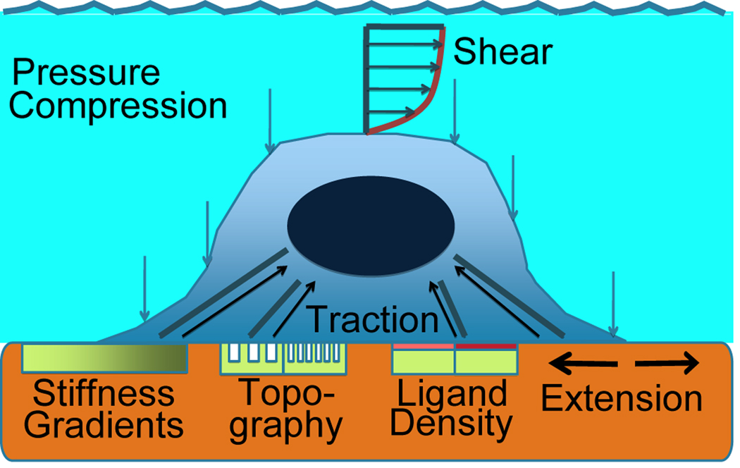
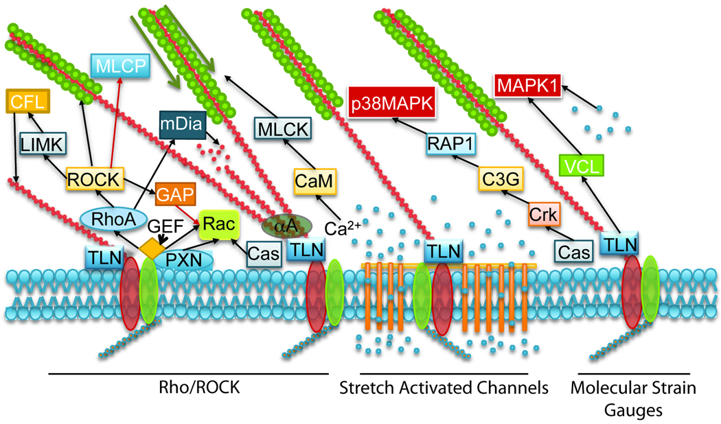
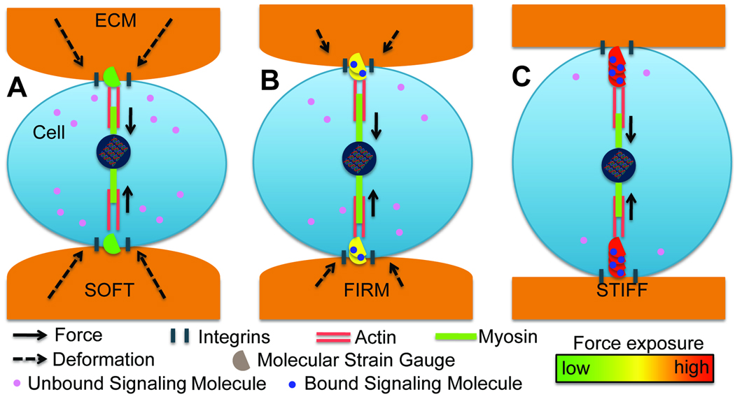
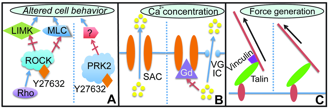
The ability of cells to extract biophysical information from their extracellular environment and convert it to biochemical signals is known as mechanotransduction. Here we detail three passive, 'inside-out' mechanotransduction mechanisms with an emphasis on the mechanosensing pathways involved in creating these signal: Rho/ROCK, stretch-activated channels, and 'Molecular Strain Gauges.' We also examine how molecular tools have been used to perturb these pathways to better understand their interconnectivity. However, perturbing pathways may have unintended confounding effects, which must also be addressed. By discovering and understanding mechanosensitive pathways, the ability to influence them for clinical applications increases.


Figures

Figure 1
Figure 1. The physiological physical microenvironment of the cell
Outside-in forces are depicted acting on the cell. On the other hand, inside-out transduction of cellular contractility can be affected by changes in stiffness gradients, ECM topography, and ligand density.
Figure 2
Figure 2. Signaling events of three major candidate mechanosensing pathways
To the left, the Rho/ROCK system is summarized, showing the mechanisms for actin nucleation, assembly, and stabilization, as well as myosin contractility. A positive feedback loop results in increasing cellular contractility, visually verified by the robust amount of stress fibers present at focal adhesions of cells on stiff microenvironments. In the center, a network of mechanosensing ion channels or “stretch activated channels” (SACs) is responding to membrane stretching, which forces open the channels. An increase in intracellular calcium concentration also provides positive feedback through favorable activation of MLCK, causing further increases in cellular contractility. Finally, molecular strain gauges, e.g. p130Cas and talin, are shown responding to increases in cellular contractility by exposing binding sites for Crk and vinculin, respectively. These pathways progress to the activation of two types of MAPK signalling kinases, which may influence changes in cell behavior further downstream. It is of note that the degree of interconnectivity between these paradigms of mechanotransduction, even as briefly overviewed in this figure, is high. Abbreviations: TLN- talin, Cas- p130Cas, VCL-vinculin, CaM-calmodulin, MLCK-myosin light chain kinase, MLCP-myosin light chain phosphatase, CFL-cofilin, LIMK-LIM Kinase, AA-alpha actinin, PXN- paxilin, GEF- guanidine exchange factor.
Figure 3
Figure 3. Changes in substrate stiffness result in a spectrum of linker protein force exposure
Because some focal adhesion proteins have been shown to undergo force-responsive unfolding events, it is feasible that changes in ECM stiffness may be a cause of discretized unfolding. Differences in unfolding at focal adhesions may result in modulation of downstream signalling molecules responsible for effecting changes in cell behavior. A) On soft substrates (~0–4 kPa), the force generated by cellular contractility is transferred to the ECM through linker proteins, but because of compliance of the ECM, linker proteins are not exposed to a high degree of force. B) On firm substrates (~8–15 kPa), compliance of the ECM decreases but does not disapear, resulting in an incremental increase in force exposure at focal adhesions. C) On firm substrates (~25 kPa-50 GPa) matrix compliance approaches zero, causing nearly the full force of cellular contractility to be focused on linker proteins in focal adhesions.
Figure 4
Figure 4. Confounding factors in manipulating mechanosensitive pathways
To further understand the mechanosensitive pathways of the cell, interfering with a hypothesized pathway and observing a change in mechanosensing can provide evidence for function. However, for a number of these interferences, confounding factors which may affect cell behavior may exist. A) The inhibitor Y27632 is a classic inhibitor of ROCK, and is used to study the role of the Rho/ROCK pathway on mechanosensing. However, Y27632 has also been shown to inhibit PRK2, whose role in mechanosensing is currently unclear. B) Intracellular calcium concentration has been shown to be important to substrate stiffness-based mechanosensing, with reduction in calcium ion concentration resulting in a decrease in stiffness-based differentiation. However, blocking stretch activated channels with the potent inhibitor gadolinium did not result in any changes in differentiation (unpublished), suggesting that calcium ions can enter the cell via other routes, including voltage gated ion channels. C) siRNA mediated knockdown of vinculin has been shown to reduce stiffness-based differentiation (unpublished), but if perturbing focal adhesions by knocking down constituents has an impact on cellular force generation, any change in cellular behavior may be attributed to a loss of contractility.

Similar articles

Cited by

References

    1. Chen KD, Li YS, Kim M, Li S, Yuan S, Chien S, Shyy JY. Mechanotransduction in response to shear stress. Roles of receptor tyrosine kinases, integrins, and Shc. J Biol Chem. 1999;274:18393–18400. - PubMed
    1. Ingber DE. Tensegrity: the architectural basis of cellular mechanotransduction. Annu Rev Physiol. 1997;59:575–599. - PubMed
    1. Szafranski JD, Grodzinsky AJ, Burger E, Gaschen V, Hung HH, Hunziker EB. Chondrocyte mechanotransduction: effects of compression on deformation of intracellular organelles and relevance to cellular biosynthesis. Osteoarthritis Cartilage. 2004;12:937–946. - PubMed
    1. Tschumperlin DJ, Dai GH, Maly IV, Kikuchi T, Laiho LH, McVittie AK, Haley KJ, Lilly CM, So PTC, Lauffenburger DA, et al. Mechanotransduction through growth-factor shedding into the extracellular space. Nature. 2004;429:83–86. - PMC - PubMed
    1. Pelham RJ, Jr, Wang YL. Cell locomotion and focal adhesions are regulated by the mechanical properties of the substrate. Biol Bull. 1998;194:348–349. discussion 349–350. - PubMed

MeSH terms

Substances