. 2011 Jul;31(13):2653-66.
doi: 10.1128/MCB.01358-10. Epub 2011 May 2.

Functional cooperation between the proteins Nck and ADAP is fundamental for actin reorganization

Affiliations

Functional cooperation between the proteins Nck and ADAP is fundamental for actin reorganization

Maor H Pauker et al. Mol Cell Biol. 2011 Jul.

Abstract

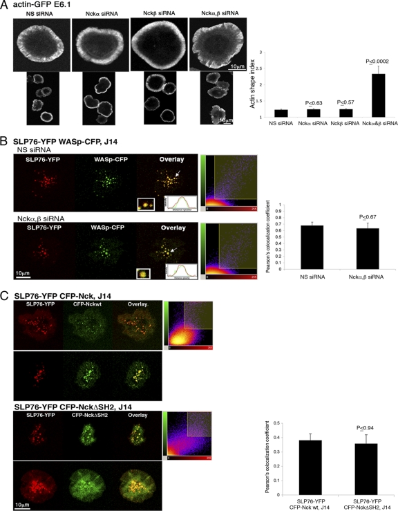
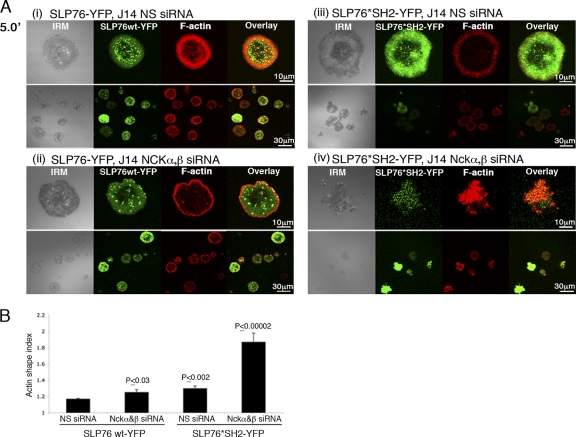
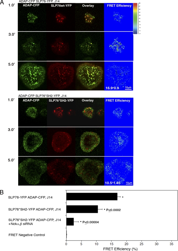
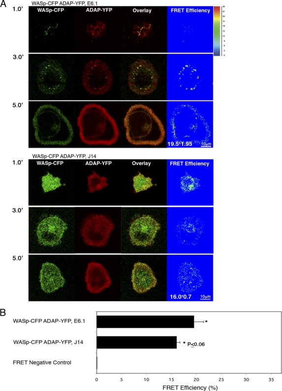
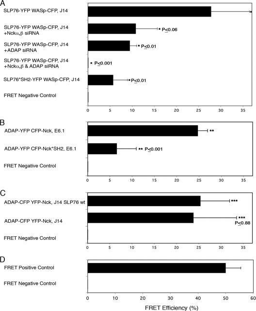
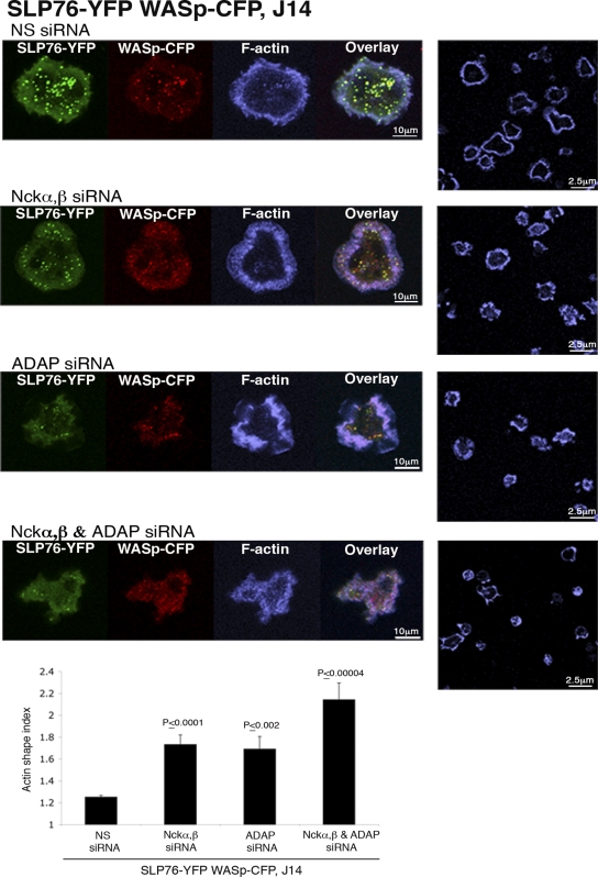
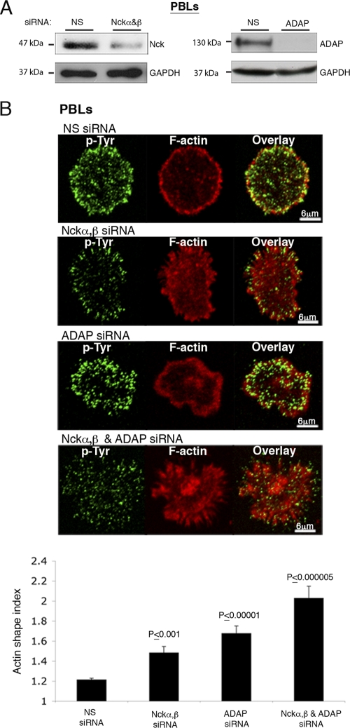
T cell antigen receptor (TCR) activation triggers profound changes in the actin cytoskeleton. In addition to controlling cellular shape and polarity, this process regulates vital T cell responses, such as T cell adhesion, motility, and proliferation. These depend on the recruitment of the signaling proteins Nck and Wiskott-Aldrich syndrome protein (WASp) to the site of TCR activation and on the functional properties of the adapter proteins linker for activation of T cells (LAT) and SH2-domain-containing leukocyte protein of 76 kDa (SLP76). We now demonstrate that Nck is necessary but insufficient for the recruitment of WASp. We show that two pathways lead to SLP76-dependent actin rearrangement. One requires the SLP76 acidic domain, crucial to association with the Nck SH2 domain, and another requires the SLP76 SH2 domain, essential for interaction with the adhesion- and degranulation-promoting adapter protein ADAP. Functional cooperation between Nck and ADAP mediates SLP76-WASp interactions and actin rearrangement. We also reveal the molecular mechanism linking ADAP to actin reorganization.


Figures

Fig. 1.
Fig. 1.
Actin polymerization and the recruitment of WASp to SLP76 signaling clusters are not solely dependent on Nck. (A) Cells were plated, fixed, and stained with phalloidin. Confocal images were collected after 5 min of activation. In the right panel, the results of >4 independent experiments are presented The actin shape index was determined as described in Materials and Methods. t tests were performed to compare the results of nonspecific (NS) siRNA treatment with the results for other experimental groups; error bars show standard errors. (B) SLP76-deficient J14 cells expressing SLP76-YFP and WASp-CFP were treated with siRNA specific to Nckα,β. Cells treated with NS siRNA served as a negative control. After 48 h, cells were plated and activated for 3 min as described for panel A. Overlays (insets) of the fluorescence peaks of the punctae are shown. Colocalization analysis is based on these fluorescence intensity peaks and was further analyzed by obtaining Pearson's colocalization coefficients (scatter diagrams). Imaging analysis was performed on more than 50 cells for each experimental group. (C) The association of Nck with SLP76 is not exclusively dependent on the Nck SH2 domain. J14 T cells expressing SLP76-YFP and the indicated CFP-Nck forms (wt or SH2 domain deletion [NckΔSH2]) were seeded on stimulatory coverslips. Live-cell images were collected 3 min after activation. Two out of 30 cells are presented for each experimental group. The combined images (overlays) are presented. A t test was performed, comparing the colocalization of CFP-Nck wt clusters with SLP76-YFP to the colocalization of the CFP-NckΔSH2 mutant with SLP76-YFP.
Fig. 2.
Fig. 2.
The SH2 domain of SLP76 is important for stabilization of SLP76 signaling clusters and for actin polymerization. (A) J14 cells expressing the SLP76 SH2 mutant form (R448K, *SH2) or SLP76 wt tagged with YFP were treated with siRNA specific to Nckα,β or NS siRNA. After 48 h, cells were plated, fixed, and stained with phalloidin 5 min (5.0′) into the spreading process (Ai to iv). IRM and combined fluorescence images (overlay) are presented. (B) The results of three independent experiments are summarized. Significant differences were found in actin rearrangement as determined by the actin shape index of SLP76 wt-YFP cells treated with NS siRNA compared to the results for the other experimental groups. A t test analysis between SLP76 wt-YFP and SLP76*SH2-YFP, treated with Nck siRNA, was performed, and the P value was found to be ≤0.00002. Error bars show standard errors.
Fig. 3.
Fig. 3.
ADAP recruitment to TCR-induced clusters is dependent on SLP76. (A) SLP76-deficient cells stably reconstituted with SLP76 wt or SLP76*SH2 tagged with YFP were transfected with ADAP-CFP. FRET analysis between SLP76-YFP and ADAP-CFP is presented. A slight but significant reduction in FRET efficiency between ADAP and SLP76*SH2 was measured in comparison to that between ADAP and SLP76 wt (P ≤ 0.0002). 1.0′, 3.0′, and 5.0′ indicate minutes of activation. (B) FRET analysis of the molecular associations between ADAP and SLP76 wt versus those between ADAP and SH2 mutant SLP76 is shown. J14 cells expressing SH2 mutant SLP76-YFP and ADAP-CFP were pretreated with specific Nck siRNAs. Cells were seeded and fixed 3 min after the initiation of spreading. The average value of FRET efficiency ± standard error obtained for each given pair is presented. The data shown are representative of >3 independent experiments.
Fig. 4.
Fig. 4.
The association between ADAP and WASp is independent of SLP76. E6.1 Jurkat T cells or SLP76-deficient J14 cells stably reconstituted with WASp-CFP were transfected with ADAP-YFP. Cells were plated, and FRET efficiency between ADAP and WASp was determined. No significant difference was measured between the two molecules in the presence versus the absence of SLP76 (P ≤ 0.06). 1.0′, 3.0′, and 5.0′ indicate minutes of activation. (B) FRET analysis of the molecular associations between WASp and ADAP. J14 or E6.1 cells expressing WASp-CFP and ADAP-YFP were seeded and were fixed 3 min after the initiation of the spreading process. The average value of FRET efficiency ± standard error obtained for a given pair is shown. The data shown are representative of >3 independent experiments.
Fig. 5.
Fig. 5.
Both Nck and ADAP participate in stabilizing the SLP76 interaction with WASp. (A) J14 cells, expressing SLP76-YFP and WASp-CFP and pretreated with specific siRNA, were seeded and fixed. The colocalization between SLP76 and WASp was observed and calculated for >3 independent experiments. FRET analysis is shown. t tests were performed comparing the results of NS siRNA treatment to the results of specific siRNA treatment for other experimental groups. (B, C) FRET analysis of the molecular associations between ADAP and Nck. Various cell types were seeded and were fixed 3 min into the spreading process. The average value of FRET efficiency ± standard error obtained for a given pair is shown. The data are representative of >3 independent experiments. (D) Results for FRET positive controls (cells expressing CFP and YFP encoded on the same plasmid, i.e., maximal FRET obtained in this system) and negative controls (CFP and YFP expressed by different plasmids and undergoing minimal FRET, i.e., only that produced by random colocalizations).
Fig. 6.
Fig. 6.
Nck and ADAP are both required for actin rearrangement. SLP76-deficient J14T cells expressing SLP76-YFP and WASp-CFP were pretreated with siRNA specific for Nckα,β and/or ADAP and were plated for 5 min on stimulatory coverslips, fixed, and stained with phalloidin (blue). Confocal images were obtained at the coverslip. The fluorescence images of SLP76-YFP, WASp-CFP, and phalloidin staining are shown. Representative fields of cells stained with phalloidin are shown for conditions in the panels on the right. The final panel shows a graph of the actin shape index obtained from the phalloidin-stained images of each sample, as explained in Materials and Methods. A graph of the results of 4 independent experiments is presented; error bars show standard errors. The results of t tests between cells treated with NS siRNA and each experimental group are presented. Significant differences were found between cells treated with Nck or ADAP siRNA and cells treated with both Nck and ADAP siRNA, with P values of ≤0.029 and ≤0.026, respectively.
Fig. 7.
Fig. 7.
Nck and ADAP are both required for actin rearrangement, as was detected in PBLs. (A) PBLs were treated with siRNA specific to Nckα,β or ADAP. After 48 h, cell lysates were prepared and analyzed for protein levels. The siRNA-mediated ADAP or Nck downregulation was compared to the level of GAPDH (glyceraldehyde-3-phosphate dehydrogenase). (B) Nckα,β and ADAP knockdown experiments of were performed with human PBLs. PBLs pretreated with siRNA specific for Nckα,β and/or ADAP were seeded on stimulatory coverslips coated with anti-CD3 and anti-CD28. After 20 min of spreading, cells were fixed and stained with phalloidin (red). Overlay images and a summary of the results of 3 independent experiments (bottom) are presented. The actin spreading index was determined as described in Materials and Methods. t tests were calculated between cells treated with NS siRNA and the experimental groups. Gene silencing of both Nckα,β and ADAP caused a dramatic impairment of actin rearrangement in comparison to that in cells treated with NS siRNA (P < 0.000005). Slight impairments were detected in cells treated with Nckα,β or ADAP siRNA (P < 0.001 and P < 0.00001, respectively). t test analyses between the results for Nck or ADAP siRNA and a combined treatment with both Nck and ADAP siRNA were performed, and the P values were <0.00061 and 0.02, respectively.

Similar articles

Cited by

References

    1. Badour K., et al. 2004. Fyn and PTP-PEST-mediated regulation of Wiskott-Aldrich syndrome protein (WASp) tyrosine phosphorylation is required for coupling T cell antigen receptor engagement to WASp effector function and T cell activation. J. Exp. Med. 199:99–112 - PMC - PubMed
    1. Badour K., et al. 2003. The Wiskott-Aldrich syndrome protein acts downstream of CD2 and the CD2AP and PSTPIP1 adaptors to promote formation of the immunological synapse. Immunity 18:141–154 - PubMed
    1. Badour K., Zhang J., Siminovitch K. A. 2004. Involvement of the Wiskott-Aldrich syndrome protein and other actin regulatory adaptors in T cell activation. Semin. Immunol. 16:395–407 - PubMed
    1. Barda-Saad M., et al. 2005. Dynamic molecular interactions linking the T cell antigen receptor to the actin cytoskeleton. Nat. Immunol. 6:80–89 - PubMed
    1. Barda-Saad M., et al. 2010. Cooperative interactions at the SLP-76 complex are critical for actin polymerization. EMBO J. 29:2315–2328 - PMC - PubMed

Publication types

MeSH terms