. 2011 Jul;31(13):2715-28.
doi: 10.1128/MCB.05151-11. Epub 2011 May 2.

The TFIIF-like Rpc37/53 dimer lies at the center of a protein network to connect TFIIIC, Bdp1, and the RNA polymerase III active center

Affiliations

The TFIIF-like Rpc37/53 dimer lies at the center of a protein network to connect TFIIIC, Bdp1, and the RNA polymerase III active center

Chih-Chien Wu et al. Mol Cell Biol. 2011 Jul.

Abstract

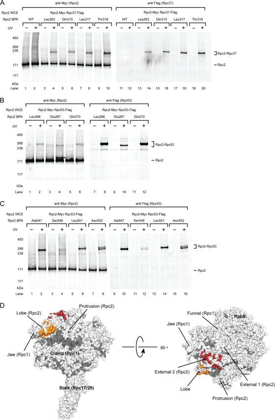
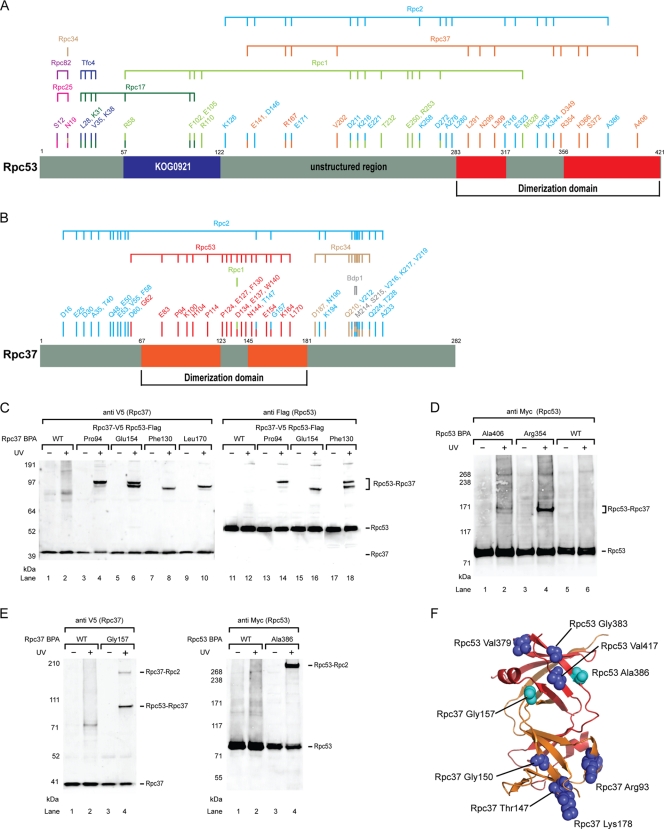
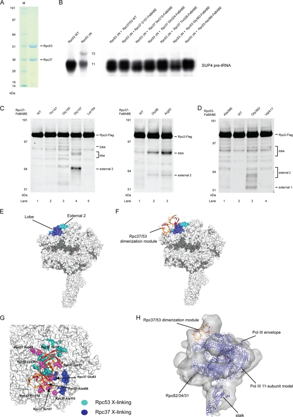
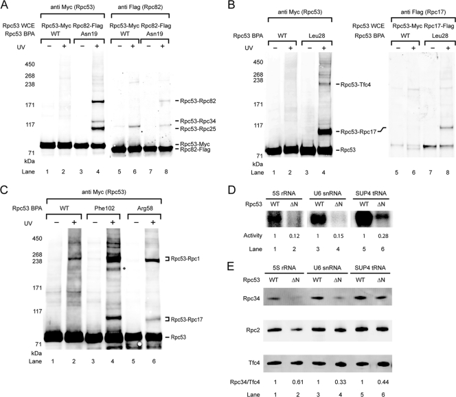
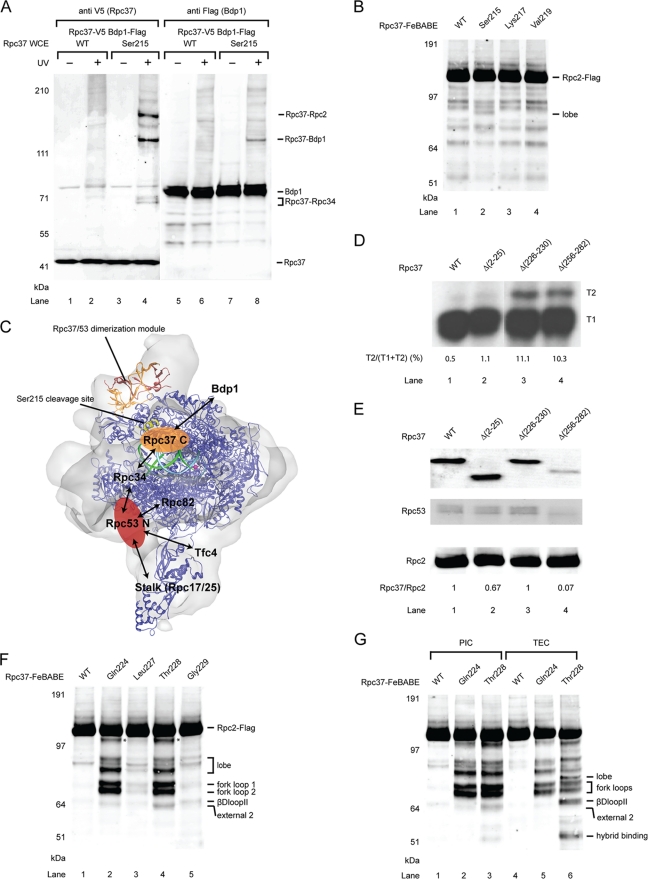
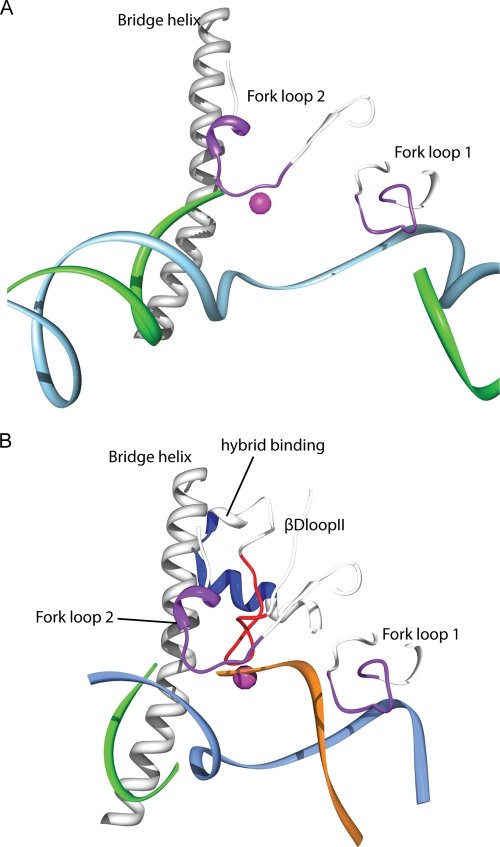
Eukaryotic RNA polymerase III (Pol III) relies on a transcription factor TFIIF-like Rpc37/53 subcomplex for promoter opening, elongation, termination, and reinitiation. By incorporating the photoreactive amino acid p-benzoyl-L-phenylalanine (BPA) into Rpc37, Rpc53, and the Rpc2 subunit of Pol III, we mapped protein-protein interactions, revealing the position of Rpc37/53 within the Pol III preinitiation complex (PIC). BPA photo-cross-linking was combined with site-directed hydroxyl radical probing to localize the Rpc37/53 dimerization module on the lobe/external 2 domains of Rpc2, in similarity to the binding of TFIIF on Pol II. N terminal to the dimerization domain, Rpc53 binds the Pol III-specific subunits Rpc82 and Rpc34, the Pol III stalk, and the assembly factor TFIIIC, essential for PIC formation. The C-terminal domain of Rpc37 interacts extensively with Rpc2 and Rpc34 and contains binding sites for initiation factor Bdp1. We also located the C-terminal domain of Rpc37 within the Pol III active center in the ternary elongation complex, where it likely functions in accurate termination. Our work explains how the Rpc37/53 dimer is anchored on the Pol III core and acts as a hub to integrate a protein network for initiation and termination.


Figures

Fig. 1.
Fig. 1.
Photo-cross-linking maps Rpc37/53 on Rpc2 lobe and external 2 domains within the Pol III PIC. (A) Western analysis of Rpc2 cross-links to Rpc37 on the lobe domain. The BPA-substituted Rpc2 residues are indicated above the lanes. Cross-links were visualized by probing with anti-Myc antibody (left panel). The identity of fusion bands was verified by probing with Flag-tagged Rpc37 (right panel). Rpc2 WCE, Rpc2 whole-cell extract. UV + or −, with or without UV irradiation. WT, wild-type Rpc2 without BPA incorporation. (B) Representative cross-links on Rpc2 lobe to Rpc53. Rpc2 and cross-links were visualized by probing with anti-Myc antibody on the left panel. The identity of the cross-linked polypeptide was verified by probing with Flag-tagged Rpc53 (right panel). Asterisk, unidentified cross-linked polypeptide. (C) Representative cross-links on Rpc2 external 2 domain to Rpc53. (D) Molecular model of the Pol III 11-subunit core. The model was built using Modeler on the basis of the homology model of Pol II core (PBD 1Y1V) and the X-ray structure of Rpc25/17 (PDB 2CKZ) (32, 35). Rpc2-Rpc37 cross-links are shown in orange and Rpc2-Rpc53 cross-links are shown in red. The magenta sphere represents magnesium at the active center.
Fig. 2.
Fig. 2.
Photo-cross-links of Rpc37/53. (A) Schematic representation of BPA incorporation in Rpc53 and cross-linked polypeptides. The region of Rpc53 homologous to the dimerization domain of human TFIIF small subunit RAP30 is shown in red. Numbers labeled above the schematic bars represent BPA-substituted residues generating cross-links. Color coding of each residue corresponds to the identity of cross-linked polypep-tide as indicated above the horizontal connecting lines. (B) Schematic representation of BPA incorporation in Rpc37 and cross-linked polypeptides. The region of Rpc37 homologous to the dimerization of human TFIIF large subunit Rap74 is shown in orange. Like in panel A, BPA-substituted residues and the corresponding cross-linked polypeptides are indicated. (C) Representative Rpc37-Rpc53 cross-links in the Rpc37 dimerization domain. Positions of BPA incorporation in Rpc37 are indicated. Anti-V5 antibody was used to reveal C-terminally V5-tagged Rpc37 and its cross-linking fusion bands (left panel). Rpc37-Rpc53 cross-links were verified by Flag epitope tagging at the C terminus of Rpc53 and probing with anti-Flag antibody (right panel). WT, wild-type without BPA incorporation. (D) Representative Rpc53-Rpc37 cross-links in the Rpc53 dimerization domain. C-terminally 13Myc-tagged Rpc53 and its cross-links are shown. Rpc53-Rpc37 cross-links were verified by Flag-epitope tagging in Rpc37 and probing with anti-Flag antibody (data not shown). (E) Rpc37- and Rpc53-Rpc2 cross-links in the Rpc37/53 dimerization domain (Rpc37 Gly157 and Rpc53 Ala386). (F) Structural model of the Rpc37/53 dimerization module, with Rpc37 shown in orange and Rpc53 shown in red. Light blue spheres indicate residues Ala386 in Rpc53 and Gly157 in Rpc37 that were shown to contact Rpc2 by photo-cross-linking. These two residues and additional residues (blue spheres) of the dimerization domain were subjected to single-cysteine substitution for FeBABE-conjugation to probe their interaction with Rpc2.
Fig. 3.
Fig. 3.
Directed hydroxyl radical probing localizes Rpc37/53 dimerization module on Rpc2 lobe/external 2. (A) SDS-PAGE analysis and Coomassie staining of the purified recombinant Rpc37/53 dimer. (B) Representative transcription assays of Rpc37/53-FeBABE derivatives. Whole-cell extracts (Rpc53 WT or ΔN) and the added Rpc37/53-FeBABE derivatives are indicated above. Each derivative in transcription assay with Rpc53 ΔN, deletion of Ser2-Leu280, whole-cell extract was shown to restore full transcription level and termination property on the SUP4 tRNA gene as the wild type extract. (C) Western analysis of hydroxyl radical cleavage of Rpc2 by FeBABE conjugated to Rpc37 surface residues in its dimerization domain. Rpc2 and peptide fragments generated from hydroxyl radical cleavage were visualized by probing with anti-Flag antibody against the C-terminally Flag epitope-tagged Rpc2. The single cysteine substitutions used for FeBABE conjugation in Rpc37 are indicated on the top, and the positions of the substituted residues are displayed in the dimerization domain model in Fig. 2F. Rpc2-Flag and the corresponding sites of cleavage fragments are indicated on the right. (D) Rpc2 cleavage from Rpc53-FeBABE derivatives in the dimerization domain. (E) Hydroxyl radical cleavage sites on the Rpc2 surface from tethered FeBABE in the Rpc37/53 dimerization domain. Calculated cleavage sites are mapped on the Pol III 11-subunit model. Based on the accuracy of cleavage site calculation, individual cleavage sites are represented by 11-residue patches centered at the cut sites and are colored in blue and cyan for cleavages from Rpc37- and Rpc53-FeBABE derivatives, respectively. (F) Model of the Rpc37/53 dimerization module on the Pol III 11-subunit core. The model was built by applying FeBABE cleavage patterns as structural restraints for ZDOCK modeling. Ribbon model represents the dimerization module, with Rpc37 in orange and Rpc53 in red. Same as in E, the hydroxyl radical cleavage sites in Rpc2 from FeBABE derivatives are colored. (G) Cross-validation of the Rpc37/53 dimerization domain-Pol III core model by the photo-cross-linking pattern from Rpc2-BPA. Blue and cyan residues on the Pol III 11-subunit (core) surface model indicate BPA-substitutions in the Rpc2 lobe/external 2 domains that produce cross-links to Rpc37 and Rpc53, respectively. The Rpc37/Rpc53 dimerization domain is shown as ribbons with Rpc37 in orange and Rpc53 in red. Magenta spheres represent BPA-substituted residues that are predicted from our model of Rpc37/53 dimerization domain on the Pol III core (shown as the white molecular surface below) to face away from Rpc2. None of these outward-facing residues in the dimerization domain cross-links to Rpc2. (H) Fit of the Rpc37/53 dimerization module-Pol III core model into the cryo-EM envelope of Pol III (25). The Pol III core is shown in blue ribbons. Rpc37 and Rpc53 are shown in orange and red, respectively.
Fig. 4.
Fig. 4.
The N-terminal region of Rpc53 contacts multiple Pol III subunits and TFIIIC for Pol III recruitment. (A) Western blot analysis showing that Rpc53 cross-links with the Rpc82/34/31 subcomplex. Rpc53 Asn19-BPA cross-linking was visualized by probing with anti-Myc antibody in the left panel. The cross-linked polypeptide Rpc82 was validated by Flag tagging Rpc82 as indicated in the right panel (anti Flag). Rpc34 and Rpc25 cross-links were also separately verified by Flag tagging (data not shown). (B) Rpc53 cross-links with the stalk protein Rpc17 and the Tfc4 subunit of the transcription factor TFIIIC. Rpc53 Leu28-BPA cross-linking was visualized by probing with anti-Myc antibody in the left panel. Right panel: the shift of Rpc53-Rpc17 fusion band was detected after Flag-tagging Rpc17 (compare lane 4 and 8), and Rpc17 in the cross-linking band was identified by probing with anti-Flag antibody (lane 8). Similarly, the Rpc53-Tfc4 cross-link in the upper fusion band was verified by Flag tagging Tfc4 (data not shown). (C) Representative Western analysis showing that BPA substitutions at Rpc53 residues Phe102 and Arg58 cross-link to Rpc1 and Rpc17 simultaneously. Asterisk, unidentified cross-linked polypeptide. (D) Rpc53 Δ(11–40) mutant compromises transcription activity. The autoradiogram shows transcription activities from the indicated Pol III promoters after PIC formation on the immobilized DNA templates and subsequent addition of NTPs spiked with 32P-labeled GTP. The relative activity of WT and Rpc53 N-terminal (aa 11 to 40) deletion (ΔN) mutant is listed below each lane. (E) The Rpc53 Δ(11–40) mutant affects PIC formation. Immobilized assay was conducted using Rpc53 mutant or Rpc53 WT whole-cell extracts with three types of Pol III promoters. Anti-Tfc4 and anti-Rpc34 antibodies were used to quantify Tfc4 and Rpc34 by Western analysis. The relative intensity of Rpc34, normalized to Tfc4, is listed below each lane. Additionally, Rpc2 was visualized by probing with anti-Flag antibody against C-terminally Flag tagged Rpc2 in the whole-cell extract.
Fig. 5.
Fig. 5.
The C-terminal region of Rpc37 contains binding sites for Bdp1 and lies near the active center for accurate termination. (A) Cross-links of Rpc37 Ser215-BPA to Bdp1, Rpc2 and Rpc34 identified by Western analysis. Left panel: Rpc37 and its cross-links were detected using anti-V5 antibody (lanes 1 to 4). Right panel: Identification of the cross-linked polypeptide Bdp1 (lanes 5 to 8) was achieved by immunostaining of the Flag tag containing Bdp1 with anti-Flag antibody. Similarly, verification of Rpc37-Rpc2 cross-link was based on probing with anti-Flag antibody for epitope tagged target (data not shown). The Rpc37-Rpc34 cross-link was identified by probing with anti-Rpc34 antibody (data not shown). (B) Position of Rpc37 Ser215 is mapped to the Rpc2 lobe. Western analysis showing Ser215-FeBABE cleaved Rpc2 lobe. The calculated cleavage site is shown in panel C. (C) Localization of the C-terminal domain of Rpc37 (Rpc37 C). Pol III 11-subunit core (blue ribbon) and Rpc37/53 dimerization module (orange and red ribbons) are fitted in Pol III cryo-EM envelope as in Fig. 2F. The yellow ribbon indicates the cleavage site in Rpc2 from the Rpc37 Ser215-FeBABE derivative. The orange oval marks the approximate position of Rpc37 C on the Pol III core. Protein-protein interactions with Bdp1 and Rpc34 are indicated. Template (cyan) and nontemplate DNA (green) are located as observed in the Pol II elongation complex (1Y1V) (35). The magenta sphere shows the active center of Pol III. Possible location of the N-terminal domain of Rpc53 (Rpc53 N; red oval) on the Pol III core is also indicated. Protein-protein interactions with Rpc34, Rpc82, Rpc17, Rpc25, and Tfc4 for Rpc53 N are marked by arrows. (D) Rpc37 C-terminal truncation mutants reduce termination efficiency. Transcription assay was conducted with whole-cell extracts containing indicated Rpc37 mutants following the immobilized template assay on the SUP4 tRNA gene that contains two consecutive T-stretches at the 3′-end of its coding sequence. T1 and T2 transcription products observed in the autoradiogram are generated from termination at the upstream and downstream T stretches, respectively. Mutants with reduced termination efficiency shift termination to site T2, whereas WT is observed to terminate mostly at T1. The relative termination shift, calculated as intensity ratio of T2 over the sum of T1 and T2, is listed below each lane. (E) Rpc37 Δ(226–230) mutant does not affect Rpc37 association with Rpc2. Coimmunoprecipitation was conducted with anti-Flag agarose gel to pull down Flag-tagged Rpc2, and probed for coimmunoprecipiated V5 epitope tagged WT or indicated Rpc37 mutants with anti-V5 antibody. The relative intensity of Rpc37 WT/mutant immunostaining is normalized to Rpc2 and listed below each lane. In addition, coimmunoprecipitated Rpc53 was visualized by probing with anti-Rpc53 antibody. In contrast to the observed coimmunoprecipitation of Rpc37 Δ(226–230) mutant, the association of Rpc37 Δ(256–282) mutant with Rpc2 was severely compromised. Coimmunoprecipitation of Rpc53 correlates with the association of Rpc37 WT/mutant with Rpc2. (F) The C-terminal region of Rpc37 is located near the Pol III active center. FeBABE hydroxyl radical cleavage of Rpc2-Flag by Rpc37-FeBABE derivatives is shown in the Western blot. Amino acid positions of Rpc37-FeBABE derivatives are listed above, and the corresponding Rpc2 cleavage sites for individual Flag epitope containing fragments are indicated on the right. The asterisk indicates an unmapped Rpc2 fragment. (G) The C-terminal region of Rpc37 moves closer to the βDloop2 of Pol III active site in the elongation complex. Rpc2 cleavages from Rpc37-FeBABE derivatives within the Pol III PIC (lanes 1 to 3) and the Pol III TEC (lanes 4 to 6) are shown.
Fig. 6.
Fig. 6.
Rpc37 influences transcription termination by interacting with fork loops and βDloopII. (A) Model of the Pol III active center and residues cleaved by Rpc37 Thr288-FeBABE derivative within Pol III PIC. The Rpc1 bridge helix and Rpc2 fork loops 1 and 2 are shown as white ribbons, and the calculated cleavage sites are shown as 11-residue purple patches. Template (cyan) and nontemplate DNA (green) strands are as observed in 1Y1V (35) by superimposition of Pol III 11-subunit model with Pol II structure. (B) Rpc37 Thr228-FeBABE cleaves βDloopII within Pol III TEC. The bridge helix, fork loops 1 and 2, βDloopII and the helix in hybrid binding domain are shown as white ribbons and the cleavage sites are indicated in red on βDloopII and in blue on hybrid binding domain, respectively. Template, nontemplate DNA and nascent RNA are as observed in 3HOW (49) and are shown as cyan, green and orange ribbons, respectively.

References

    1. Bartholomew B., Durkovich D., Kassavetis G. A., Geiduschek E. P. 1993. Orientation and topography of RNA polymerase III in transcription complexes. Mol. Cell. Biol. 13:942–952 - PMC - PubMed
    1. Bartholomew B., Kassavetis G. A., Geiduschek E. P. 1991. Two components of Saccharomyces cerevisiae transcription factor IIIB (TFIIIB) are stereospecifically located upstream of a tRNA gene and interact with the second-largest subunit of TFIIIC. Mol. Cell. Biol. 11:5181–5189 - PMC - PubMed
    1. Brachmann C. B., et al. 1998. Designer deletion strains derived from Saccharomyces cerevisiae S288C: a useful set of strains and plasmids for PCR-mediated gene disruption and other applications. Yeast 14:115–132 - PubMed
    1. Braun B. R., Bartholomew B., Kassavetis G. A., Geiduschek E. P. 1992. Topography of transcription factor complexes on the Saccharomyces cerevisiae 5 S RNA gene. J. Mol. Biol. 228:1063–1077 - PubMed
    1. Brow D. A., Guthrie C. 1990. Transcription of a yeast U6 snRNA gene requires a polymerase III promoter element in a novel position. Genes Dev. 4:1345–1356 - PubMed

Publication types

MeSH terms

Substances

LinkOut - more resources