. 2011 Jul;31(13):2552-65.
doi: 10.1128/MCB.05045-11. Epub 2011 May 2.

Oncogenic tyrosine kinases target Dok-1 for ubiquitin-mediated proteasomal degradation to promote cell transformation

Affiliations

Oncogenic tyrosine kinases target Dok-1 for ubiquitin-mediated proteasomal degradation to promote cell transformation

Justyna A Janas et al. Mol Cell Biol. 2011 Jul.

Abstract

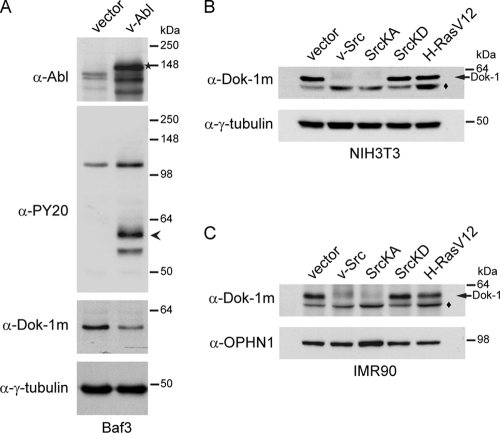
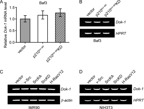
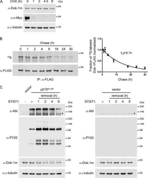
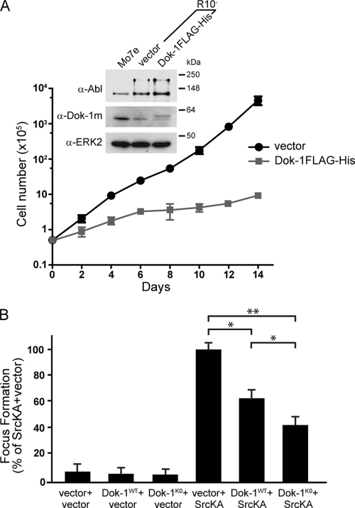
Cellular transformation induced by oncogenic tyrosine kinases is a multistep process involving activation of growth-promoting signaling pathways and inactivation of suppressor molecules. Dok-1 is an adaptor protein that acts as a negative regulator of tyrosine kinase-initiated signaling and opposes oncogenic tyrosine kinase-mediated cell transformation. Findings that its loss facilitates transformation induced by oncogenic tyrosine kinases suggest that Dok-1 inactivation could constitute an intermediate step in oncogenesis driven by these oncoproteins. However, whether Dok-1 is subject to regulation by oncogenic tyrosine kinases remained unknown. In this study, we show that oncogenic tyrosine kinases, including p210(bcr-abl) and oncogenic forms of Src, downregulate Dok-1 by targeting it for degradation through the ubiquitin-proteasome pathway. This process is dependent on the tyrosine kinase activity of the oncoproteins and is mediated primarily by lysine-dependent polyubiquitination of Dok-1. Importantly, restoration of Dok-1 levels strongly suppresses transformation of cells expressing oncogenic tyrosine kinases, and this suppression is more pronounced in the context of a Dok-1 mutant that is largely refractory to oncogenic tyrosine kinase-induced degradation. Our findings suggest that proteasome-mediated downregulation of Dok-1 is a key mechanism by which oncogenic tyrosine kinases overcome the inhibitory effect of Dok-1 on cellular transformation and tumor progression.


Figures

FIG. 1.
FIG. 1.
p210bcr-abl induces downregulation of Dok-1 protein levels in a tyrosine kinase activity-dependent manner. (A, B) Mo7e cells (A) and Baf3 cells (B) were stably transduced with empty control vector or a p210bcr-abl-, p210bcr-ablKD-, or p210bcr-ablT315I-expressing retroviral vector. Where indicated, the cells were cultured in the presence of 1 μM STI571. Tyrosine phosphorylation of cellular proteins was determined in total cell lysates by Western blot analysis with antiphosphotyrosine (α-PY20) Ab. The blots were reprobed with anti-Abl Ab, a polyclonal Ab raised against the PH domain of Dok-1 (α-Dok-1PH), and anti-γ-tubulin Ab as a loading control. The arrowhead indicates the position of tyrosine-phosphorylated Dok-1, as confirmed by anti-Dok-1m immunoprecipitation (IP), followed by Western blot analysis with antiphosphotyrosine (α-PY20) and anti-Dok-1PH Abs (B, right). HC, Ig heavy chain. (C) Total cell lysates prepared from Mo7e cells stably transduced with empty control vector or a p210bcr-abl- or p210bcr-abl KD-expressing retroviral vector were analyzed by Western blotting with anti-Abl Ab and a monoclonal anti-Dok-1 (α-Dok-1m) Ab. The blots were reprobed with anti-Dok-1PH Ab and anti-γ-tubulin Ab as a loading control. (D) Baf3/TonB210.1 cells expressing p210bcr-abl under the control of the tetracycline-inducible promoter were treated with increasing concentrations of doxycycline to induce p210bcr-abl expression. Total cell lysates were analyzed by Western blotting with antiphosphotyrosine (α-PY20) Ab, anti-Abl Ab, anti-Dok-1m Ab, and anti-γ-tubulin Ab as a loading control. (E) Total cell lysates prepared from Mo7e cells and K562 cells treated with the indicated concentrations of STI571, or a control vehicle, were analyzed by Western blotting with antiphosphotyrosine (α-PY20) Ab, anti-Abl Ab, anti-Dok-1PH Ab, and anti-γ-tubulin Ab as a loading control. Asterisks indicate the positions of endogenous c-Abl.
FIG. 2.
FIG. 2.
v-Abl and oncogenic Src kinases, but not H-RasV12, induce Dok-1 downregulation. (A) Baf3 cells were stably transduced with empty control vector or v-Abl-expressing retroviral vector. Total cell lysates were analyzed by immunoblotting with antiphosphotyrosine (α-PY20) Ab, anti-Abl Ab, anti-Dok-1m Ab, and anti-γ-tubulin Ab as a loading control. The arrowhead indicates the position of tyrosine-phosphorylated Dok-1. The asterisk indicates the position of v-Abl. (B, C) NIH 3T3 cells (B) and IMR90 cells (C) were stably transduced with empty control vector or a v-Src-, SrcKA-, SrcKD-, or H-RasV12-expressing retroviral vector. Total cell lysates were analyzed by Western blotting with anti-Dok-1m Ab and anti-γ-tubulin Ab (B) or anti-OPHN1 Ab (C) as loading controls. The diamonds denote nonspecific bands.
FIG. 3.
FIG. 3.
Oncogenic tyrosine kinases do not affect the levels of Dok-1 mRNA. (A, B) Total RNA was isolated from Baf3 cells stably transduced with empty control vector or a p210bcr-abl- or p210bcr-ablKD-expressing retroviral vector, and the levels of Dok-1 transcripts were measured by Q-PCR (A) and RT-PCR (B) using gene-specific primers as described in Materials and Methods. The hypoxanthine-guanine phosphoribosyltransferase (HPRT) gene was amplified as an internal control. (C, D) RT-PCR analysis of Dok-1 mRNA levels in IMR90 cells (C) and NIH 3T3 cells (D) stably transduced with the indicated retroviral vectors. β-actin (C) and HPRT (D) genes were used as internal controls.
FIG. 4.
FIG. 4.
p210bcr-abl downregulates Dok-1 by reducing Dok-1 protein stability. (A) Baf3 cells were treated with protein synthesis inhibitor cycloheximide (CHX; 200 μg/ml). At indicated time points, cells were lysed, and total cell lysates were analyzed by Western blotting for Dok-1, c-Myc, and γ-tubulin as a loading control. (B) Half-life of Dok-1 in Baf3 cells. Baf3 cells stably expressing FLAG-tagged Dok-1 were pulse-labeled with [35S]methionine/cysteine for 1 h, chased in fresh medium, and harvested at the indicated times postchase. Cell lysates were subjected to anti-FLAG immunoprecipitation (IP), and the immunoprecipitates were resolved by SDS-PAGE followed by blotting (left). 35S-labeled Dok-1FLAG was quantified by phosphorimaging, and total levels of Dok-1FLAG protein were quantified by Western blotting with anti-FLAG Ab. The fraction of radiolabeled Dok-1FLAG remaining is shown for each chase time point (right). Error bars represent the standard deviations (SD) from three independent experiments. (C) Baf3 cells transduced with an empty control vector or p210bcr-abl-expressing retroviral vector were cultured in the absence (−) or presence (+) of 1 μM STI571. The STI571-treated p210bcr-abl-expressing cells (left) or control vector-expressing cells (right) were lysed at 0 to 8 h after STI571 removal, and total cell lysates were analyzed by Western blotting with antiphosphotyrosine (α-PY20) Ab, anti-Abl Ab, anti-Dok-1m Ab, and anti-γ-tubulin Ab as a loading control. Asterisks indicate the positions of endogenous c-Abl.
FIG. 5.
FIG. 5.
Oncogenic tyrosine kinase-induced downregulation of Dok-1 requires proteasome activity. (A) Baf3 cells transduced with p210bcr-abl or empty control vector were treated with proteasome inhibitor MG132 (10 μM) for the indicated times. Total lysates were analyzed by Western blotting using antiphosphotyrosine (α-PY20) Ab, anti-Abl Ab, anti-Dok1PH Ab, and anti-γ-tubulin Ab as a loading control. The asterisk indicates the position of endogenous c-Abl. (B, C) IMR90 cells stably transduced with empty control vector or a v-Src-, SrcKA-, SrcKD-, or H-RasV12-expressing retroviral vector were treated with proteasome inhibitors MG132 (10 μM), ALLN (50 μM), or control vehicle for 8 h. Total protein and RNA were isolated. (B) The lysates were analyzed by Western blotting with antiphosphotyrosine (α-PY20) Ab, anti-Dok-1m Ab, and anti-γ-tubulin Ab as a loading control. The diamonds denote nonspecific bands. (C) The levels of Dok-1 transcript were measured by RT-PCR, as described for Fig. 3C. (D) IMR90 cells stably transduced with empty control vector or a v-Src- or SrcKA-expressing retroviral vector were treated with proteasome inhibitors, as described for panel B. Total cell lysates were analyzed by Western blotting using anti-Src Ab and anti-γ-tubulin Ab as a loading control.
FIG. 6.
FIG. 6.
p210bcr-abl and oncogenic forms of Src kinase induce polyubiquitination of Dok-1. (A) HEK293 cells stably transduced with FLAG-tagged Dok-1 (HEK293/Dok-1FLAG) were transfected with a His6-tagged ubiquitin (Ub)-encoding vector, or empty control vector, together with either a vector encoding v-Src or empty control vector. A total of 36 h later, the cells were treated with MG132 (10 μM) or control vehicle for 8 h. Ubiquitin conjugates were affinity precipitated under highly denaturing conditions by Ni-NTA chelate chromatography, resolved by SDS-PAGE, and analyzed by immunoblotting with anti-FLAG Ab. The right panel is a lighter exposure of data shown in the left panel. (B, C) HEK293/Dok-1FLAG cells were transiently transfected with empty control vector or His6-tagged ubiquitin-expressing vector and with increasing amounts of expression vectors encoding SrcKA, SrcKD, or empty vector control (B) or with vectors encoding p210bcr-abl, p210bcr-ablKD, or empty control vector (C), as indicated. Cells were lysed 48 h after transfection, and the lysates were subjected to Ni-NTA affinity precipitation and Western blotting with anti-FLAG Ab. The diamond denotes a nonspecific band.
FIG. 7.
FIG. 7.
Role of lysine residues in oncogenic tyrosine kinase-induced ubiquitination and downregulation of Dok-1. (A, B) HEK293T cells were transiently cotransfected with the indicated expression vectors. A total of 36 h posttransfection, cells were left untreated (A) or were treated with MG132 (10 μM) or control vehicle for 8 h (B). Ubiquitin conjugates were affinity precipitated under denaturing conditions by Ni-NTA chelate chromatography and analyzed by Western blotting with anti-HA Ab. Diamonds denote nonspecific bands. (C) NIH 3T3 cells stably expressing HA-tagged wild-type Dok-1 (Dok-1WTHA), lysineless Dok-1 mutant (Dok-1K0HA), or empty control vector were transduced with a retroviral vector expressing SrcKA or an empty control vector. Total cell lysates were subjected to Western blot analysis with anti-HA Ab and anti-γ-tubulin Ab as a loading control. (Left) A representative example from three independent experiments is shown. (Right) Quantification of Dok-1WTHA and Dok-1K0HA protein levels. Data were normalized to γ-tubulin and then to the value of 1.0 for the non-SrcKA-expressing condition. Error bars represent SD. (D) Lysates prepared from cells described for panel C were subjected to anti-HA immunoprecipitation (IP), followed by Western blotting with antiphosphotyrosine (α-PY20) and anti-HA Abs. (E) Lysates prepared from cells described for panel C were left untreated or were treated with calf intestinal phosphatase (CIP) as described in Materials and Methods, and subjected to Western blotting with antiphosphotyrosine (α-PY20) Ab, anti-HA Ab, and anti-γ-tubulin Ab as a loading control. The arrowhead indicates the position of the tyrosine-phosphorylated Dok-1HA proteins. (F) NIH 3T3 cells stably transduced with Dok-1WTHA, Dok-1K0HA, or control empty vector were serum starved and were either left untreated (−) or treated (+) with PDGF (12.5 ng/ml) for 10 min. Cell extracts were subjected to anti-HA immunoprecipitation (IP). A portion of the final eluates (6%) was subjected to Western blotting with anti-HA Ab (bottom), and the remaining eluted immune complexes were analyzed by Western blotting with anti-RasGAP Ab. HC, Ig heavy chain. (G) HEK293T cells transfected with the indicated expression vectors were serum starved and stimulated with EGF (50 ng/ml) for 10 min. Cell extracts were subjected to anti-FLAG immunoprecipitation (IP), followed by Western blotting with anti-HA and anti-FLAG Abs.
FIG. 8.
FIG. 8.
Restoration of Dok-1 expression levels suppresses cellular proliferation and transformation induced by p210bcr-abl and oncogenic Src kinases. (A) Growth curve of R10 cells stably transduced with a Dok-1FLAG-His-expressing retroviral vector or empty control vector. The number of cells was plotted on a logarithmic scale against time on a linear scale. The data represent the means ± SD from triplicate experiments. Data from a representative of three independent experiments are shown. (Inset) Total cell lysates prepared from Mo7e cells and R10 cells expressing Dok-1FLAG-His or empty control vector were analyzed by Western blotting with anti-Abl, anti-Dok-1m Ab, and anti-ERK2 Ab as a loading control. (B) Focus formation assay. NIH 3T3 cells stably transduced with Dok-1WTHA-, Dok-1K0HA-expressing retroviral vector, or empty control vector were infected with a SrcKA-expressing retroviral vector or an empty control vector. Focus-forming activities are presented as percentages of the SrcKA-plus-vector condition. Data represent the means ± SD from three independent experiments performed in triplicate. *, P < 0.05; **, P < 0.01 versus SrcKA plus vector by t test.

References

    1. Abram C. L., Courtneidge S. A. 2000. Src family tyrosine kinases and growth factor signaling. Exp. Cell Res. 254:1–13 - PubMed
    1. Bache K. G., Slagsvold T., Stenmark H. 2004. Defective downregulation of receptor tyrosine kinases in cancer. EMBO J. 23:2707–2712 - PMC - PubMed
    1. Barnes D. J., Schultheis B., Adedeji S., Melo J. V. 2005. Dose-dependent effects of Bcr-Abl in cell line models of different stages of chronic myeloid leukemia. Oncogene 24:6432–6440 - PubMed
    1. Berger A. H., et al. 2010. Identification of DOK genes as lung tumor suppressors. Nat. Genet. 42:216–223 - PMC - PubMed
    1. Berman E., et al. 2000. Characterization of two novel sublines established from a human megakaryoblastic leukemia cell line transfected with p210(BCR-ABL). Leuk. Res. 24:289–297 - PubMed

Publication types

MeSH terms