. 2011 Jul 1;286(26):23296-307.
doi: 10.1074/jbc.M111.237586. Epub 2011 May 2.

Survivin monomer plays an essential role in apoptosis regulation

Affiliations

Survivin monomer plays an essential role in apoptosis regulation

Marat S Pavlyukov et al. J Biol Chem. .

Abstract

Survivin was initially described as an inhibitor of apoptosis and attracted growing attention as one of the most tumor-specific genes in the human genome and a promising target for cancer therapy. Lately, it has been shown that survivin is a multifunctional protein that takes part in several crucial cell processes. At first, it was supposed that survivin functions only as a homodimer, but now data indicate that many processes require monomeric survivin. Moreover, recent studies reveal a special mechanism regulating the balance between monomeric and dimeric forms of the protein. In this paper we studied the mutant form of survivin that was unable to dimerize and investigated its role in apoptosis. We showed that survivin monomer interacts with Smac/DIABLO and X-linked inhibitor of apoptosis protein (XIAP) both in vitro and in vivo. Due to this feature, it protects cells from caspase-dependent apoptosis even more efficiently than the wild-type survivin. We also identified that mutant monomeric survivin prevents apoptosis-inducing factor release from the mitochondrial intermembrane space, protecting human fibrosarcoma HT1080 cells from caspase-independent apoptosis. On the other hand, our results indicate that only wild-type survivin, but not the monomer mutant form, enhances tubulin stability in cells. These findings suggest that survivin partly performs its functions as a monomer and partly as a dimer. The mechanism of dimer-monomer balance regulation may also work as a "switcher" between survivin functions and thereby explain remarkable functional diversities of this protein.


Figures
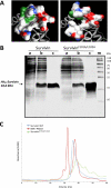
FIGURE 1.
FIGURE 1.
Wild-type survivin and survivinF101A/L102A structures. A, model of dimerization region electrostatic surface of WT survivin (left) (25) (Protein Data Bank ID code 1E31) and survivinF101A/L102A (right). Positively charged areas are shown in blue, negatively charged regions are in red, and 101 and 102 residues are marked in green. B, Coomassie Blue-stained SDS PAGE: m, molecular mass marker; a, lysates from E. coli cells containing vector pQE80-survivin or pQE80-survivinF101A/L102A; b, lysates from E. coli cells containing vector pQE80-survivin or pQE80-survivinF101A/L102A after isopropyl 1-thio-β-d-galactopyranoside induction; c, recombinant survivin or survivinF101A/L102A purified from E. coli strain BL-21(DE3). C, gel filtration profile for purified His-tagged recombinant WT survivin (blue line) or survivinF101A/L102A (green line). Protein molecular mass markers are shown in red: BSA (66 kDa) and RNaseA (14 kDa).
FIGURE 2.
FIGURE 2.
Survivin interaction with Smac/DIABLO. A, interaction of survivin and survivinF101A/L102A with Smac/DIABLO and Δ63Smac/DIABLO. Recombinant mature Smac/DIABLO-His6 (Smac) and Δ63Smac/DIABLO-His6 (Δ63Smac) were incubated with recombinant WT His6-survivin (survivin) or His6-survivinF101A/L102A (survivinF101A/L102A), immobilized on protein G-Sepharose with antibodies against survivin or with nonimmune antibodies as a negative control (---). Immunoblotting with primary antibodies against Smac/DIABLO was used to detect bound proteins. B, fluorescence images of CFP, YFP, and FRET emission in HT1080 cells co-transfected with pTagCFP-N-Smac/DIABLO and pEYFP-C-survivin plasmids. Scale bars, 10 μm. C, acceptor photobleaching on YFP-survivin or YFP-survivinF101A/L102A and CFP, Smac/DIABLO-CFP or Δ63Smac/DIABLO-CFP co-expressing cells. YFP was selectively photobleached at 514 nm. The graph shows the CFP fluorescence intensity versus acceptor photobleaching time. D, same as in C for HT1080 cells co-expressing fragmented survivin: YFP-survivin 1–109 or YFP-survivin 84–109 and Smac/DIABLO-CFP. The data represent three independent experiments.
FIGURE 3.
FIGURE 3.
Interaction between survivin and XIAP. A, co-immunoprecipitation of XIAP from CFP-survivin-, CFP-survivinF101A/L102A-, or CFP-survivinD53A-expressing cells. HT1080 cells were transformed with pTagCFP-C-survivin, pTagCFP-C-survivinF101A/L102A or pTagCFP-C-survivinD53A plasmids. Then, cells were lysed, and endogenous XIAP was immunoprecipitated (IP) with antibodies against XIAP. Precipitated proteins were detected by immunoblotting (W) with primary antibodies against XIAP or survivin. B, acceptor photobleaching on YFP-survivin or YFP-survivinF101A/L102A and CFP-XIAP co-expressing cells. YFP was selectively photobleached at 514 nm. The graph shows the CFP fluorescence intensity versus acceptor photobleaching time. The data represent three independent experiments.
FIGURE 4.
FIGURE 4.
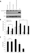
Effect of different survivin mutants on caspase-dependent apoptosis in HT1080 cells. A, HT1080 cells were infected by lentiviral vector encoding WT survivin (WT), survivinF101A/L102A (F101A/L102A), survivinD53A (D53A), survivinD53A/F101A/L102A (D53A/F101A/L102A), or CFP. Cells were grown on selective puromycin-containing medium. Lysates from cells expressing WT survivin, survivin mutant forms, or CFP were analyzed by immunoblotting with primary antibodies against CFP. Blotting with primary antibodies against actin was used as a control for equivalent protein loading. B, noninfected HT1080 cells or cells expressing WT survivin (WT), survivinF101A/L102A (F101A/L102A), survivinD53A (D53A), survivinD53A/F101A/L102A (D53A/F101A/L102A), or CFP (CFP) were treated with 50 μm cisplatin (black bars) or left untreated (gray bars). After 48 h of incubation both attached and detached cells were fixed and permeabilized. To estimate the apoptosis level, permeabilized cells were stained with primary antibodies against cleaved caspase-3 and secondary antibodies conjugated with Alexa Fluor 488, then cells were analyzed on FACScan flow cytometer. The figures represent means ± S.D. (error bars) from three independent experiments (*, p < 0.05). C, relative Bcl-XL mRNA levels in HT1080 cells expressing WT survivin (WT), survivinF101A/L102A (F101A/L102A), survivinD53A (D53A), survivinD53A/F101A/L102A (D53A/F101A/L102A), or CFP (CFP). RNA was purified, reverse-transcribed into cDNA, and quantitative RT-PCR was performed with primers to human Bcl-XL. The data are mean ± S.D. for triplicate experiments (**, p > 0.5).
FIGURE 5.
FIGURE 5.
Effect of Smac/DIABLO knockdown on caspase-dependent cisplatin-induced apoptosis in HT1080 cells expressing survivin mutants. A, siRNA knockdown Smac/DIABLO. HT1080 cells expressing WT survivin (WT), survivinF101A/L102A (F101A/L102A), survivinD53A (D53A), survivinD53A/F101A/L102A (D53A/F101A/L102A), or CFP (CFP) were transfected with control siRNA (Luc) or siRNA against Smac/DIABLO (Smac). After a 72-h incubation, Smac/DIABLO expression was analyzed with immunoblotting. B, HT1080 cells expressing WT survivin, survivinF101A/L102A (F101A/L102A), survivinD53A (D53A), survivinD53A/F101A/L102A (D53A/F101A/L102A), or CFP (CFP) were transfected with control siRNA (gray bars) or siRNA against Smac/DIABLO (black bars). After a 24-h incubation cells were treated with 50 μm cisplatin (CP). After an additional 48-h incubation the level of caspase-dependent apoptosis was determined as in Fig. 4B. The figures represent mean values ± S.D. (error bars) from three independent experiments (*, p < 0.05; **, p > 0.1).
FIGURE 6.
FIGURE 6.
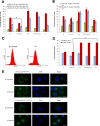
Effect of different survivin mutants on cisplatin-induced caspase-independent apoptosis in HT1080 cells. A, HT1080 cells expressing WT survivin (WT), survivinF101A/L102A (F101A/L102A), survivinD53A (D53A), survivinD53A/F101A/L102A (D53A/F101A/L102A), or CFP (CFP) were treated with 50 μm cisplatin (CP) in the presence (red bars) or absence (blue bars) of 20 μm Z-VAD-fmk. Cells treated with 20 μm Z-VAD-fmk without cisplatin were taken as a negative control (green bars). After a 48-h incubation apoptosis level was determined by staining with annexin-PE and flow cytometry. The figures represent mean values ± S.D. (error bars) from three independent experiments (*, p < 0.05). B, same as in A for cells treated with 0.5 μm staurosporine (STS). C, HT1080 cells expressing CFP treated with 50 μm cisplatin (right) or left untreated (left). After a 48-h incubation the level of AIF released from IMS was measured according to the method described above. D, same as C for HT1080 cells expressing WT survivin (WT), survivinF101A/L102A (F101A/L102A), survivinD53A (D53A), survivinD53A/F101A/L102A (D53A/F101A/L102A), or CFP (CFP). The figures represent mean values ± S.D. from two independent experiments (*, p < 0.05). E, HT1080 cells expressing survivinF101A/L102A or survivinD53A/F101A/L102A treated with 100 μm cisplatin or left untreated. After a 48-h incubation cells were fixed, immunostained with antibodies against AIF, and treated with DAPI. Images were taken using a Nikon DIAPHOT 300 fluorescent microscope. Scale bars, 20 μm.
FIGURE 7.
FIGURE 7.
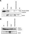
Effect of survivin mutants on microtubule stability. A, HT1080 cells expressing WT survivin (WT), survivinF101A/L102A (F101A/L102A), survivinD53A (D53A), survivinD53A/F101A/L102A (D53A/F101A/L102A), or CFP (CFP) were lysed and analyzed by immunoblotting with primary antibodies against acetylated α-tubulin. Blotting with primary antibodies against actin was used as a control for equivalent protein loading. B, HT1080 cells expressing WT survivin (WT), survivinF101A/L102A (F101A/L102A), survivinD53A (D53A), or survivinD53A/F101A/L102A (D53A/F101A/L102A) were lysed in microtubule stabilization buffer, and polymerized tubulin was separated from soluble tubulin by centrifugation. Pellet was analyzed by immunoblotting with primary antibodies against α-tubulin, and supernatants were analyzed by immunoblotting with primary antibodies against actin.
FIGURE 8.
FIGURE 8.
Proposed model depicting different function of dimeric and monomeric forms of survivin in cells. Survivin (ribbon representation; Protein Data Bank ID code 1E31) appears to be in dynamic equilibrium between dimeric and monomeric forms, and the balance can be regulated via posttranslational modifications. Survivin monomer prevents cells from caspase-dependent and caspase-independent apoptosis and is a part of the chromosome passenger complex during mitosis. Survivin dimer is required for microtubule stability and takes part in STAT3-dependent repression of transcription.

References

    1. Ambrosini G., Adida C., Altieri D. C. (1997) Nat. Med. 3, 917–921 - PubMed
    1. Velculescu V. E., Madden S. L., Zhang L., Lash A. E., Yu J., Rago C., Lal A., Wang C. J., Beaudry G. A., Ciriello K. M., Cook B. P., Dufault M. R., Ferguson A. T., Gao Y., He T. C., Hermeking H., Hiraldo S. K., Hwang P. M., Lopez M. A., Luderer H. F., Mathews B., Petroziello J. M., Polyak K., Zawel L., Kinzler K. W. (1999) Nat. Genet. 23, 387–388 - PubMed
    1. Altieri D. C. (2008) Nat. Rev. Cancer 8, 61–70 - PubMed
    1. Delacour-Larose M., Thi M. N., Dimitrov S., Molla A. (2007) Cell Cycle 6, 1878–1885 - PMC - PubMed
    1. Lens S. M., Medema R. H. (2003) Cell Cycle 2, 507–510 - PubMed

Publication types

MeSH terms