. 2011 May 1;25(9):984-95.
doi: 10.1101/gad.1998611.

Phosphoinositide [PI(3,5)P2] lipid-dependent regulation of the general transcriptional regulator Tup1

Affiliations

Phosphoinositide [PI(3,5)P2] lipid-dependent regulation of the general transcriptional regulator Tup1

Bong-Kwan Han et al. Genes Dev. .

Abstract

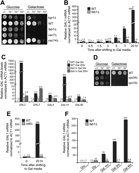
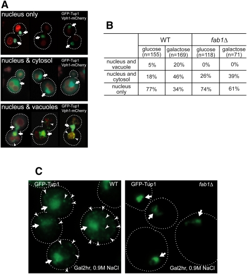
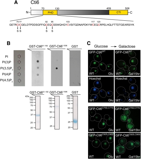
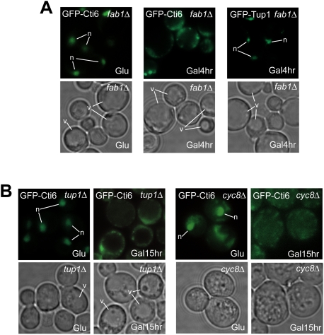
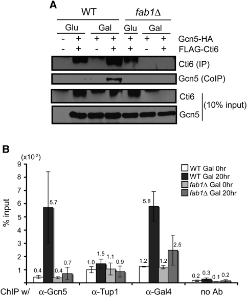
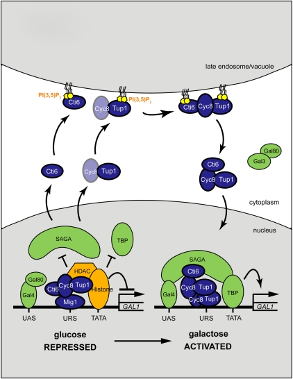
Transcriptional activity of a gene is governed by transcriptional regulatory complexes that assemble/disassemble on the gene and control the chromatin architecture. How cytoplasmic components influence the assembly/disassembly of transcriptional regulatory complexes is poorly understood. Here we report that the budding yeast Saccharomyces cerevisiae has a chromatin architecture-modulating mechanism that is dependent on the endosomal lipid PI(3,5)P(2). We identified Tup1 and Cti6 as new, highly specific PI(3,5)P(2) interactors. Tup1--which associates with multiple transcriptional regulators, including the HDAC (histone deacetylase) and SAGA complexes--plays a crucial role in determining an activated or repressed chromatin state of numerous genes, including GAL1. We show that, in the context that the Gal4 activation pathway is compromised, PI(3,5)P(2) plays an essential role in converting the Tup1-driven repressed chromatin structure into a SAGA-containing activated chromatin structure at the GAL1 promoter. Biochemical and cell biological experiments suggest that PI(3,5)P(2) recruits Cti6 and the Cyc8-Tup1 corepressor complex to the late endosomal/vacuolar membrane and mediates the assembly of a Cti6-Cyc8-Tup1 coactivator complex that functions to recruit the SAGA complex to the GAL1 promoter. Our findings provide important insights toward understanding how the chromatin architecture and epigenetic status of a gene are regulated by cytoplasmic components.


Figures

Figure 1.
Figure 1.
Tup1 binds PI(3,5)P2 lipid with a high specificity. (A, top panel) GST-Tup1 and GST-Atg18 bound PI(3,5)P2 with a high specificity, while GST-PHPLCδ specifically bound PI(4,5)P2. GST protein alone did not bind any PI lipid. (Bottom panel) Equivalent amount of purified, bacterially expressed GST fusion proteins (50 μg) were used for protein–lipid overlay assay. (B) Vph1-mCherry marks the limiting membrane of vacuoles (red). A pool of GFP-Tup1 localized at the vacuolar membrane (arrowhead), in addition to the nuclear Tup1 (arrow), was observed in some wild-type (WT) cells (see Supplemental Fig. S2B). In contrast, the vacuolar localization of Tup1 was not observed in fab1Δ cells.
Figure 2.
Figure 2.
PI(3,5)P2 and Cti6 are required for transcriptional induction of GAL1. (A) SEY6210 fab1Δ and vac7Δ cells exhibited the Gal phenotype. SEY6210 vac14Δ cells exhibited the Gal+ phenotype. SEY6210 tup1Δ cells showed slower Gal growth than wild-type (WT) cells. (B) RT-qPCR analysis showed gradual induction of GAL1 mRNA in SEY6210 wild-type cells. At 20 h after Gal shift, a 490-fold induction of GAL1 mRNA was observed in wild-type cells. No detectable increase in GAL1 mRNA was observed in fab1Δ cells at 20 h after Gal shift. The results that are shown with a standard deviation are from three independent analyses. (C) The mRNA levels of GAL2 and GAL10 were highly induced in SEY6210 wild-type cells, but remained constitutively repressed in SEY6210 fab1Δ cells at 20 h after Gal shift. The mRNA levels of GAL3, GAL4, and GAL10 were induced a few-fold in SEY6210 wild-type cells, while, in fab1Δ cells, the levels of GAL4 and GAL80 mRNA remained repressed (see a caveat in the text for the GAL3 mRNA results of fab1Δ cells). (D) SEY6210 vps34Δ and vps15Δ cells, which produce no PI(3)P and PI(3,5)P2, exhibited the Gal phenotype. (E) SEY6210 cti6Δ cells exhibited a severe defect in GAL1 mRNA induction. (F) SEY6210 cells grown in Glu medium were shifted to Gly medium and grown for several hours to derepress GAL genes. When shifted to Gal medium, GAL1 mRNA was induced very rapidly in wild-type cells, whereas GAL1 remained constitutively repressed in fab1Δ cells.
Figure 3.
Figure 3.
Gal and hyperosmotic stress redistribute Tup1. (A) Montages of cells containing nuclear GFP-Tup1 only (top panel), nuclear and cytosolic GFP-Tup1 (middle panel), or nuclear and vacuolar GFP-Tup1 (bottom panel) and Vph1-mCherry. Nuclear GFP-Tup1 is indicated by an arrow and vacuolar membrane GFP-Tup1 is marked by an arrowhead. (B) Cells grown in Glu or Gal (2 h after shift) medium were imaged, analyzed, and scored for one of the three categories of GFP-Tup1 localization (nuclear only, nuclear and cytosolic, or nuclear and vacuolar membrane). Gal medium significantly enhanced cytosolic and vacuolar membrane localization of Tup1 in wild-type (WT) cells. In contrast, although Gal medium increased cytosolic localization of Tup1 in fab1Δ cells, vacuolar membrane localization of Tup1 was not observed. (C) When wild-type cells in Gal medium were treated with 0.9 M NaCl, Tup1 formed puncta (arrowhead) in the cytoplasm in most of the wild-type cells in addition to the nuclear Tup1 (arrow), whereas Tup1 did not form cytoplasmic puncta in fab1Δ cells.
Figure 4.
Figure 4.
Cti6 binds PI(3,5)P2 lipid, and the Cys residues in Cti6's PHD domain are important for nuclear localization of Cti6 in Gal medium. (A) Cti6 contains a PHD domain and a CTI domain. The PHD domain contains several Cys residues (red) that are potentially involved in zinc binding and important for PIP binding. Two Cti6 mutants were generated, such as Cti6C75S,C77S (Cys75–Cys77 → Ser75–Ser77) and Cti6C92S,C95S (Cys92–Cys95 → Ser92–Ser95). (B, top panel) Full-length Cti6 bound PI(3,5)P2 with high specificity, while the region (amino acids 51∼150) encompassing the PHD domain of Cti6 bound PI(3)P, PI(3,5)P2, and PI(4)P. (Bottom panel) Bacterially expressed GST fusion proteins were purified, and the same amounts of proteins were used for the protein–lipid overlay assay. (C) Wild-type Cti6 localized primarily in the nucleus in wild-type (WT) cells in both Glu and Gal media. The Cti6 mutants Cti6C75S,C77S and Cti6C92S,C95S localized in the nucleus in wild-type cells in Glu medium, but a significant portion of the mutant Cti6 proteins accumulated in the cytoplasm upon Gal shift.
Figure 5.
Figure 5.
PI(3,5)P2 and the Cyc8–Tup1 complex are required for nuclear localization of Cti6 in Gal medium. (A) Cti6 localized primarily in the nucleus in fab1Δ cells in Glu medium, but Cti6 began to accumulate in the cytoplasm upon Gal shift, and a significant portion of Cti6 was observed in the cytoplasm at 4 h after Gal shift. Tup1 localized primarily in the nucleus at 4 h after Gal shift. (B) Cti6 localized in the nucleus in tup1Δ cells in Glu medium, but began to accumulate in the cytoplasm upon Gal shift. Similarly, in cyc8Δ cells, Cti6 localized in the nucleus in Glu medium, but a significant portion of Cti6 localized in the cytoplasm in Gal medium. Also, a significant portion of Cti6 often localized in the cytoplasm of cyc8Δ cells in Glu medium.
Figure 6.
Figure 6.
PI(3,5)P2 is required for Cti6's interaction with SAGA and for the recruitment of the SAGA complex to the GAL1 promoter in Gal medium. (A) Flag-tagged Cti6 was immunoprecipitated from the extracts prepared from the same number of cells grown in Glu and Gal media. Coimmunoprecipitated Gcn5 significantly increased from wild-type (WT) cell extract in Gal medium compared with in Glu medium. In contrast, coimmunoprecipitated Gcn5 was not detectable from fab1Δ cell extracts in both Glu and Gal media. Much less Cti6 was immunoprecipitated from fab1Δ cells in Gal medium, probably due to its instability in the cell extract (see the text). (B) ChIP-qPCR experiments were performed with wild-type and fab1Δ cells containing HA-tagged Gcn5 protein (n = 3). The amount of Gcn5 associated with the GAL1 promoter increased significantly (∼15-fold) in wild-type cells in Gal medium. In contrast, no significant increase of Gcn5 was detected in fab1Δ cells in Gal medium. The amount of Tup1 associated with the GAL1 promoter was not significantly changed in both wild-type and fab1Δ cells in Gal medium compared with Glu medium. A slightly increased amount of Tup1 associated with the GAL1 promoter was detected in wild-type cells in Gal medium. The amount of Gal4 bound to the GAL1 promoter increased a few-fold in wild-type cells (about fivefold) and fab1Δ cells (about twofold) in Gal medium.
Figure 7.
Figure 7.
A model for the PI(3,5)P2 function in GAL induction. We propose that (1) upon Gal shift, there is dynamic nucleocytoplasmic shuttling of Cti6, Cyc8, and Tup1; (2) PI(3,5)P2 recruits Cyc8–Tup1 and Cti6 to the late endosomal/vacuolar membrane and mediates the assembly of the Cti6–Cyc8–Tup1 complex that is required for Cti6's shuttling into the nucleus; and (3) once in the nucleus, the Cti6–Cyc8–Tup1 complex functions as a transcriptional coactivator by recruiting the SAGA complex to the GAL1 promoter.

Similar articles

Cited by

References

    1. Abrajano JJ, Qureshi IA, Gokhan S, Molero AE, Zheng D, Bergman A, Mehler MF 2010. Corepressor for element-1-silencing transcription factor preferentially mediates gene networks underlying neural stem cell fate decisions. Proc Natl Acad Sci 107: 16685–16690 - PMC - PubMed
    1. Audhya A, Emr SD 2003. Regulation of PI4,5P2 synthesis by nuclear-cytoplasmic shuttling of the Mss4 lipid kinase. EMBO J 22: 4223–4236 - PMC - PubMed
    1. Ballas N, Mandel G 2005. The many faces of REST oversee epigenetic programming of neuronal genes. Curr Opin Neurobiol 15: 500–506 - PubMed
    1. Bhaumik SR, Green MR 2001. SAGA is an essential in vivo target of the yeast acidic activator Gal4p. Genes Dev 15: 1935–1945 - PMC - PubMed
    1. Bonangelino CJ, Nau JJ, Duex JE, Brinkman M, Wurmser AE, Gary JD, Emr SD, Weisman LS 2002. Osmotic stress-induced increase of phosphatidylinositol 3,5-bisphosphate requires Vac14p, an activator of the lipid kinase Fab1p. J Cell Biol 156: 1015–1028 - PMC - PubMed

MeSH terms