. 2011 May 9;208(5):909-21.
doi: 10.1084/jem.20102518. Epub 2011 May 2.

Trypanosoma cruzi subverts the sphingomyelinase-mediated plasma membrane repair pathway for cell invasion

Affiliations

Trypanosoma cruzi subverts the sphingomyelinase-mediated plasma membrane repair pathway for cell invasion

Maria Cecilia Fernandes et al. J Exp Med. .

Abstract

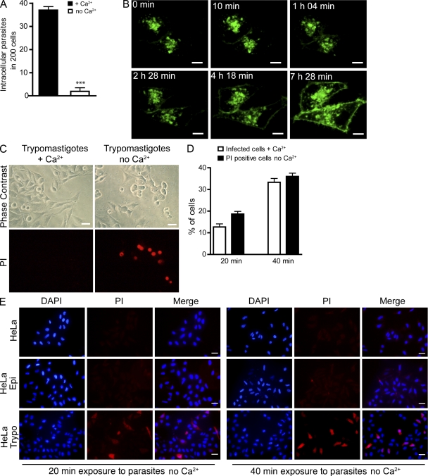
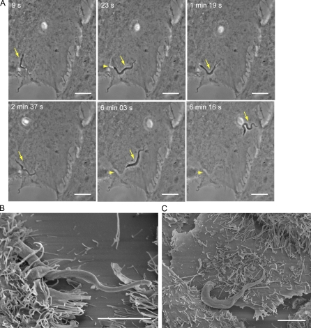
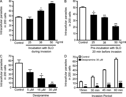
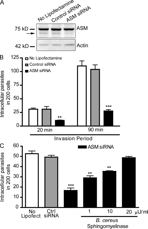
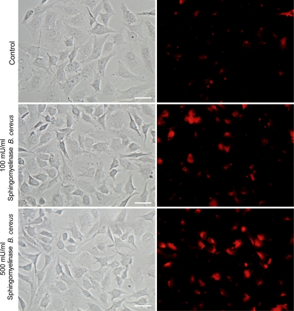
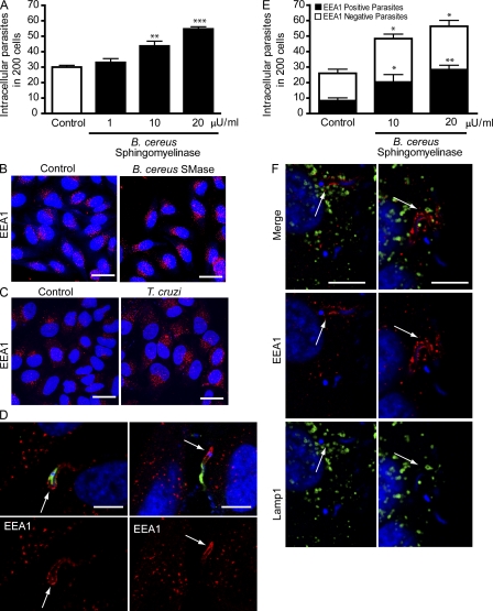
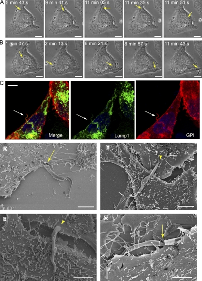
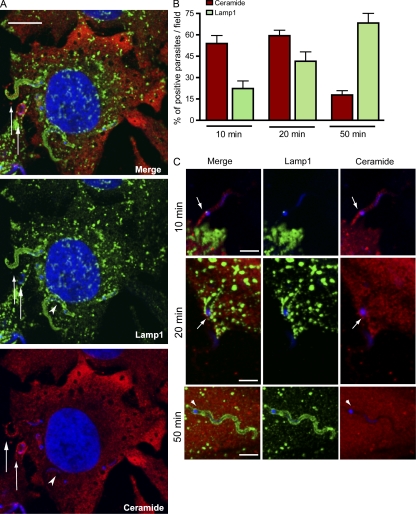
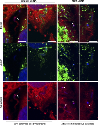
Upon host cell contact, the protozoan parasite Trypanosoma cruzi triggers cytosolic Ca(2+) transients that induce exocytosis of lysosomes, a process required for cell invasion. However, the exact mechanism by which lysosomal exocytosis mediates T. cruzi internalization remains unclear. We show that host cell entry by T. cruzi mimics a process of plasma membrane injury and repair that involves Ca(2+)-dependent exocytosis of lysosomes, delivery of acid sphingomyelinase (ASM) to the outer leaflet of the plasma membrane, and a rapid form of endocytosis that internalizes membrane lesions. Host cells incubated with T. cruzi trypomastigotes are transiently wounded, show increased levels of endocytosis, and become more susceptible to infection when injured with pore-forming toxins. Inhibition or depletion of lysosomal ASM, which blocks plasma membrane repair, markedly reduces the susceptibility of host cells to T. cruzi invasion. Notably, extracellular addition of sphingomyelinase stimulates host cell endocytosis, enhances T. cruzi invasion, and restores normal invasion levels in ASM-depleted cells. Ceramide, the product of sphingomyelin hydrolysis, is detected in newly formed parasitophorous vacuoles containing trypomastigotes but not in the few parasite-containing vacuoles formed in ASM-depleted cells. Thus, T. cruzi subverts the ASM-dependent ceramide-enriched endosomes that function in plasma membrane repair to infect host cells.


Figures

Figure 1.
Figure 1.
T. cruzi trypomastigotes wound mammalian cells and trigger the Ca2+-dependent plasma membrane repair response mediated by lysosomal exocytosis. (A) HeLa cells were incubated with trypomastigotes in the presence (membrane repair condition) or absence (nonrepair condition) of Ca2+ for 40 min, fixed, and stained for microscopic quantification of intracellular parasites. The data correspond to the mean of triplicates ± SD. ***, P < 0.0001, Student’s t test. (B) Time-lapse live images of cells transduced with Lamp1-RFP and incubated with trypomastigotes in the presence of Ca2+. The images show a single confocal plane of cells expressing Lamp1-RFP (green) during interaction with trypomastigotes. The first time point (0 min) was acquired after 20 min incubation of cells with 108 trypomastigotes. Bars, 10 µm. (Video1.) (C) Cell permeabilization and PI (red) influx in response to trypomastigotes in the absence (nonrepair condition) or presence (repair condition) of Ca2+. Live cells were imaged by phase contrast and epifluorescence 40 min after trypomastigote addition. Bars, 20 µm. (D) Quantification of the percentage of PI-positive cells (in Ca2+ free medium) in comparison with the percentage of cells infected with trypomastigotes (in Ca2+-containing medium). Trypomastigotes were allowed to invade cells for the indicated time periods and fixed coverslips were examined to determine the percentage of infected cells or PI-positive cells. Error bars show mean of triplicates ± SD. (E) Fixed cells after incubation with noninfective epimastigotes (Epi) or trypomastigotes (Trypo) in Ca2+-free medium in the presence of PI (red). Cell nuclei were stained with DAPI (blue). Bars, 20 µm. These results are representative of three independent experiments.
Figure 2.
Figure 2.
Interaction of T. cruzi trypomastigotes with host cells before invasion causes mechanical deformations on the plasma membrane. (A) Time-lapse phase-contrast live images of trypomastigotes interacting with a HeLa cell (Video 2). The gliding movement of attached trypomastigotes (arrow) cause reversible plasma membrane deformations (arrowhead). Bars, 10 µm. (B and C) Scanning electron microscopy images of trypomastigotes during early stages (10 min) of interaction with host cells. Trypomastigotes are observed gliding under cells (B) or in close contact with the plasma membrane at the cell periphery (C). Bars, 5 µm. These results are representative of two independent experiments.
Figure 3.
Figure 3.
Host cell wounding and repair modulates T. cruzi invasion. (A) HeLa cells were exposed to trypomastigotes in the absence (white column) or presence (black columns) of increasing concentrations of SLO for 20 min, fixed, and processed for quantification of intracellular parasites. The data correspond to the mean of triplicates ± SD. *, P = 0.0149, Student’s t test comparing control and 38 ng/ml SLO-treated cells; ***, P < 0.0001, Student’s t test comparing control and 50 ng/ml SLO-treated cells. (B) Cells were pretreated for 20 min with increasing concentrations of SLO, washed, further incubated for 20 min, and exposed to trypomastigotes for 20 min followed by fixation and quantification of intracellular parasites. The data correspond to the mean of triplicates ± SD. *, P = 0.0182, Student’s t test comparing control and 25 ng/ml SLO-treated cells; ***, P < 0.0006, Student’s t test comparing control and 38 ng/ml or 50 ng/ml SLO-treated cells. (C) HeLa cells were treated with the indicated concentrations of the ASM inhibitor desipramine for 1 h, washed and exposed to trypomastigotes for 30 min, fixed, and processed for determination of the number of intracellular parasites. The data correspond to the mean of triplicates ± SD. *, P = 0.0125, Student’s t test; **, P = 0.0016, Student’s t test comparing control (white bars) and treated (black bars) bars. (D) Cells preincubated with 30 µM desipramine for 1 h were exposed to trypomastigotes for the indicated periods of time, washed, and processed for quantification of intracellular parasites. The data correspond to the mean of triplicates ± SD. **, P < 0.0014, Student’s t test; ***, P < 0.0001, Student’s t test comparing control (white bars) and treated (black bars) cells. These results are representative of four independent experiments.
Figure 4.
Figure 4.
RNAi-mediated silencing of lysosomal ASM inhibits host cell invasion by T. cruzi, and exposure to extracellular sphingomyelinase restores parasite entry. (A) Immunoblot with anti-ASM antibodies in extracts of HeLa cells treated with control or ASM siRNA. The arrow points to the band corresponding to ASM; the higher additional band is an unspecific reaction. Immunoblot was probed with anti-actin antibodies for loading control. (B) Quantification of trypomastigote invasion in siRNA-treated HeLa cells. After siRNA treatment, cells were incubated with trypomastigotes for the indicated periods, fixed, and processed for quantification of intracellular parasites. The data correspond to the mean of triplicates ± SD. **, P = 0.0073, Student’s t test; ***, P < 0.0001, Student’s t test comparing control (gray bars) and ASM siRNA-treated cells (black bars). (C) siRNA-treated HeLa cells were incubated with trypomastigotes in the presence or absence of the indicated concentrations of bacterial sphingomyelinase for 30 min, fixed, and processed for quantification of intracellular parasites. The data correspond to the mean of triplicates ± SD. **, P < 0.0014; ***, P = 0.0003, Student’s t test comparing control (gray bars) and treated cells (black bars). These results are representative of four independent experiments.
Figure 5.
Figure 5.
Anti-ceramide monoclonal antibodies detect sphingomyelinase-induced increase in surface ceramide. Hela cells were treated with the indicated concentrations of Bacillus cereus sphingomyelinase for 30 min. After fixation with PFA, coverslips were labeled with 15B4 anti-ceramide monoclonal antibodies for 1 h followed by secondary antibodies. Ceramide labeling is shown in red. Bars, 20 µm. These results are representative of three independent experiments.
Figure 6.
Figure 6.
Exposure to T. cruzi trypomastigotes or to exogenous sphingomyelinase induces formation of EEA1-positive endosomes in host cells. (A) After exposure to trypomastigotes for 20 min, HeLa cells were fixed and processed for quantification of intracellular parasites. The data correspond to the mean of triplicates ± SD. **, P = 0.0078, Student’s t test comparing control (white bar) and 10 µU/ml sphingomyelinase–exposed cells (black bar); ***, P < 0.0001, Student’s t test comparing control and 20 µU/ml sphingomyelinase–treated cells. (B) EEA1-positive endosomes in HeLa cells incubated (SMase) or not (control) with 20 µU/ml sphingomyelinase for 20 min. DAPI DNA stain (blue), anti-EEA1 antibodies (red). Bars = 20 µm. (C) EEA1-positive endosomes in HeLa cells incubated (T. cruzi) or not (control) with trypomastigotes for 20 min. DAPI, blue; anti-EEA1 antibodies, red. Bars, 20 µm. (D) After 20 min of infection, HeLa cells were incubated with anti–T. cruzi antibodies (green) to stain extracellular parasites, followed by permeabilization and staining with anti-EEA1 (red). Arrows point to EEA1 staining associated with the intracellular region of partially internalized parasites. DAPI (blue). Bars, 10 µm. (E) Quantification of the number of intracellular trypomastigotes in EEA1 positive vacuoles 20 min after invasion in the presence of sphingomyelinase. After infection, cells were incubated with anti–T. cruzi antibodies to stain extracellular parasites, followed by permeabilization and staining with anti-EEA1. The data correspond to the mean of triplicates ± SD. *, P < 0.04, Student’s t test; **, P = 0.002, Student’s t test, comparing control and treated cells. (F) After 20 min of infection, HeLa cells were fixed, permeabilized and stained with anti-EEA1 (red) and anti-Lamp1 (green) antibodies. DAPI, blue. Arrows indicate intracellular parasites within EEA1-enriched parasitophorous vacuoles and closely associated with Lamp1-containing lysosomes. Bars, 10 µm. These results are representative of three independent experiments.
Figure 7.
Figure 7.
Recently internalized parasites are highly motile and protrude from host cells. (A) Time lapse of T. cruzi internalization and intracellular movement in a HeLa cell (Video 3). The arrow indicates the invading trypomastigote. Bars, 10 µm. (B) Time-lapse images of the same cell in A (initiated ∼15 min after the last frame of the previous video), showing that the trypomastigote remains highly motile and then protrudes from the host cell. The arrow indicates the intracellular trypomastigote. Bars, 10 µm. (C) Confocal images of a trypomastigote protruding from the host cell surface 30 min after invasion of a HeLa cell transduced with the plasma membrane marker GPI-YFP (red) and Lamp1-RFP (green). After infection, cells were fixed and DNA was stained with DAPI (blue). The arrow indicates an internalized parasite already within a Lamp1-positive compartment, protruding from the GPI-labeled host cell plasma membrane. Bar, 10 µm. (D–G) Scanning EM images of protruding trypomastigotes. HeLa cells exposed to trypomastigotes for 20 min were washed and incubated for an additional 15 min before fixation and processing for scanning electron microscopy. Bars, 5 µm. Arrowheads indicate the continuity between the plasma membrane and the protrusion, and arrows indicate sites where the plasma membrane was fractured, revealing the parasitophorous vacuole membrane. These results are representative of two independent experiments.
Figure 8.
Figure 8.
Vacuoles containing recently internalized parasites are enriched in ceramide while gradually acquiring Lamp1. (A) Single optical section of a HeLa cell expressing Lamp1-RFP (green) infected with trypomastigotes for 15 min, fixed, permeabilized, and stained with anti-ceramide antibodies (red). Trypomastigotes (arrows) can be observed in vacuoles enriched in ceramide (red), of which one has already fused with Lamp1-RFP–containing lysosomes (green). The arrowhead indicates a Lamp1-positive parasite vacuole that is negative for ceramide. Bar, 10 µm. (B) Quantification of intracellular parasites found in ceramide or Lamp1-enriched vacuoles over time. After 10 or 20 min of exposure to trypomastigotes, cells were washed and either fixed or incubated for an additional 30 min before fixation (50-min time point). Cells were then permeabilized, stained with antibodies to ceramide (red) or Lamp1 (green), and confocal Z series were obtained in 15 fields for each condition, followed by quantification of parasites associated with ceramide and Lamp1. The data represents mean ± SD of the percentage of positive parasites per field (n = 15). (C) Representative images (single optical sections) of each time point in B. At 10 and 20 min, protruding parasites were often observed in ceramide-enriched vacuoles (arrows). Ceramide, red; Lamp1, green; DAPI, blue. The arrowheads indicate a Lamp1-positive parasite vacuole that is negative for ceramide. Bars, 5 µm. These results are representative of three independent experiments.
Figure 9.
Figure 9.
Ceramide-enriched T. cruzi-containing vacuoles are dependent on ASM activity. HeLa cells treated with control or ASM siRNA were exposed to trypomastigotes for 20 min, fixed, and processed as described in Fig. 7. Confocal Z series analysis performed in 15 fields for each condition revealed a smaller percentage of ceramide positive vacuoles in cells treated with ASM siRNA (24%) when compared with control siRNA (60%). Representative images of single optical planes for each condition are shown (two examples of control siRNA and two examples of ASM-siRNA). Ceramide, red; Lamp1, green; DAPI, blue. The arrows indicate parasites within vacuoles positive for ceramide staining in cells treated with the control siRNA. Arrowheads point to the DAPI-stained kinetoplasts of trypomastigotes within vacuoles negative for ceramide staining, in cells treated with ASM siRNA. Bars, 5 µm. These results are representative of three independent experiments.

Comment in

Similar articles

Cited by

References

    1. Andrade L.O., Andrews N.W. 2004. Lysosomal fusion is essential for the retention of Trypanosoma cruzi inside host cells. J. Exp. Med. 200:1135–1143 10.1084/jem.20041408 - DOI - PMC - PubMed
    1. Andrews N.W., Whitlow M.B. 1989. Secretion by Trypanosoma cruzi of a hemolysin active at low pH. Mol. Biochem. Parasitol. 33:249–256 10.1016/0166-6851(89)90086-8 - DOI - PubMed
    1. Andrews N.W., Hong K.S., Robbins E.S., Nussenzweig V. 1987. Stage-specific surface antigens expressed during the morphogenesis of vertebrate forms of Trypanosoma cruzi. Exp. Parasitol. 64:474–484 10.1016/0014-4894(87)90062-2 - DOI - PubMed
    1. Andrews N.W., Abrams C.K., Slatin S.L., Griffiths G. 1990. A T. cruzi-secreted protein immunologically related to the complement component C9: evidence for membrane pore-forming activity at low pH. Cell. 61:1277–1287 10.1016/0092-8674(90)90692-8 - DOI - PubMed
    1. Bertello L.E., Andrews N.W., de Lederkremer R.M. 1996. Developmentally regulated expression of ceramide in Trypanosoma cruzi. Mol. Biochem. Parasitol. 79:143–151 10.1016/0166-6851(96)02645-X - DOI - PubMed

Publication types

MeSH terms