. 2011 May 9;208(5):1083-92.
doi: 10.1084/jem.20092277. Epub 2011 May 2.

Thrombospondin 1 inhibits inflammatory lymphangiogenesis by CD36 ligation on monocytes

Affiliations

Thrombospondin 1 inhibits inflammatory lymphangiogenesis by CD36 ligation on monocytes

Claus Cursiefen et al. J Exp Med. .

Abstract

Lymphangiogenesis plays an important role in tumor metastasis and transplant outcome. Here, we show that thrombospondin-1 (TSP-1), a multifunctional extracellular matrix protein and naturally occurring inhibitor of angiogenesis inhibits lymphangiogenesis in mice. Compared with wild-type mice, 6-mo-old TSP-1-deficient mice develop increased spontaneous corneal lymphangiogenesis. Similarly, in a model of inflammation-induced corneal neovascularization, young TSP-1-deficient mice develop exacerbated lymphangiogenesis, which can be reversed by topical application of recombinant human TSP-1. Such increased corneal lymphangiogenesis is also detected in mice lacking CD36, a receptor for TSP-1. In these mice, repopulation of corneal macrophages with predominantly WT mice via bone marrow reconstitution ameliorates their prolymphangiogenic phenotype. In vitro, exposure of WT macrophages to TSP-1 suppresses expression of lymphangiogenic factors vascular endothelial growth factor (VEGF)-C and VEGF-D, but not of a primarily hemangiogenic factor VEGF-A. Inhibition of VEGF-C is not detected in the absence or blockade of CD36. These findings suggest that TSP-1, by ligating CD36 on monocytic cells, acts as an endogenous inhibitor of lymphangiogenesis.


Figures

Figure 1.
Figure 1.
TSP-1−/− mice display mild isolated lymphangiogenesis. (A–D) Immunofluorescence analysis of LYVE-1+++/CD31+ lymphatic vessels (L, arrows) emanating from the limbal arcade (Li) toward the center of the cornea in 6-mo-old TSP-1−/− (A-C) or WT (control, D) mice. Blood vessels (B, arrowheads) in the limbal arcade stain intensely with anti-CD31, but not with anti–LYVE-1. (E) Electron micrograph of the corneal stromal lymphatic vessel in TSP-1−/− mice showing erythrocyte-free lumen (Lu). Arrowheads denote the thin endothelial lining with absent basement membrane and absent pericyte coverage. Inset shows higher magnification view. (F and G) WT control and TSP-1−/− complete corneal flat-mounts stained with LYVE-1 (Li, limbus, arrowhead; L, lymphatic vessel, arrow). Note isolated lymphatic vessel traversing the whole cornea in G (L, arrow). (H) Morphometric comparison demonstrates a significantly increased spontaneous corneal lymphangiogenesis in TSP-1−/− mice compared with WT controls (*, P < 0.05; n = 10; all mice aged 6 mo). (I) Morphometric quantification of lymphatic vessels in the ear skin from young (p10) or adult (p30) WT and TSP-1−/− mice (*, P < 0.05; n = 5 per group, compared with WT controls). Error bars indicate SD.
Figure 2.
Figure 2.
TSP-1 regulates the lymphangiogenic response in a mouse model of inflammation-associated corneal neovascularization. Representative segments of corneal flat mounts from WT and TSP-1−/− mice stained with CD31 are shown in green (blood vessels) and LYVE-1 is shown in red (lymphatic vessels; A; Li, Limbal vascular arcade: bottom, center of the cornea: top; B; L, lymph vessels). (C) Morphometric quantification of lymphvascularized area in WT and TSP-1−/− mice (*, P < 0.05; n = 10 per group; age 8 wk). (D) Morphometric analysis of vascularized areas with lymphatic vessels in sutured corneas of WT mice treated with three subconjunctival injections and topical eye drops (x3 daily) of recombinant human (rh) TSP-1 in comparison to control (*, P < 0.05; n = 8 per group, age 8 wk). Error bars indicate SD.
Figure 3.
Figure 3.
Increased transcription of the lymphangiogenic growth factor VEGF-C in TSP-1−/− mice. VEGF-C mRNA levels in corneas of 8-wk-old (A) and 6-mo-old (B) TSP-1−/− mice in comparison to WT controls. (C) Flow cytometric detection of CD11b+ macrophages in the corneas harvested from 6-mo-old TSP-1−/− mice (24.6%) compared with WT controls (19.7%). (D) Expression of VEGF-C in thioglycollate-elicited macrophages from TSP-1−/− mice (age 8 wk) compared with WT mice detected by real-time PCR. Corneas were pooled from five mice per group in each experiment. Expression of VEGF-C relative to the GAPDH in four PCR reactions per RNA sample is indicated. *, P < 0.05 compared with WT control. Error bars indicate SD.
Figure 4.
Figure 4.
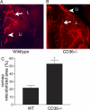
Increased inflammation-induced lymphangiogenesis in CD36−/− mice. Representative segments of corneal flat mounts from WT (A) and a CD36−/− (B) mice. (C) Morphometric comparison of lymphvascularized area between WT control and CD36−/− mice (*, P < 0.05; n = 10 per group; age 8 wk). Li, limbal vascular arcade bottom; center of the cornea, top; flat mount stained with LYVE-1, red (lymphatic vessels); L, lymph vessels. Error bars indicate SD.
Figure 5.
Figure 5.
CD36 on macrophages regulates the lymphangiogenic response in the cornea. (A) The macrophage marker CD11b (green) and the TSP-1 receptor CD36 (red) are colocalized (yellow) on monocytic cells in the normal corneal stroma. (B) The specific lymphatic marker LYVE-1 (red) is not colocalized with antibodies against CD36 (green), and is expressed by blood vessels detectable at the limbus. (C) Morphometric comparison of corneal lymphvascularized area between WT (n = 8) control and CD36−/− (n = 9) mice (reconstituted with WT bone marrow) in a mouse model of inflammatory corneal neovascularization. Error bars indicate SD.
Figure 6.
Figure 6.
TSP-1 down-regulates expression of VEGF-C in macrophages via CD36. Relative expression of VEGF-C (A, B, D, and E), VEGF-D (F), and VEGF-A (G) message and VEGF-C protein (C) in thioglycollate-elicited macrophages from WT or CD36−/− mice: (A) freshly harvested, (B–G) cultured in the absence or presence of TGFβ2, TSP-1, or CD36 blocking and control peptides. Total RNA harvested from macrophages was subjected to real-time PCR analysis to detect expression of VEGF-C, VEGF-D, and VEGF-A mRNA and to detect VEGF-C protein culture supernatants were analyzed by ELISA. Although freshly harvested CD36−/− macrophages express significantly lower levels of VEGF-C compared with WT controls (A) these macrophages retain their ability to respond to exogenously provided TGFβ2 in culture by increasing expression of VEGF-C (both message [B] and protein [C]) compared with WT controls. Inhibition of VEGF-C expression detectable in WT macrophages cultured with TSP-1 (5 µg/ml) is not detected in similarly treated CD36 −/− macrophages (D). In the presence of CD36-binding TSP-1–derived peptide capable of blocking CD36–TSP-1 interaction (CD36 blocking peptide), the inhibitory effect of TSP-1 on the VEGF-C expression in WT macrophages is significantly reversed (E). Treatment of WT macrophages to TSP-1 also inhibited the expression of the lymphangiogenesis factor VEGF-D (F), but not that of predominantly hemangiogenic factor VEGF-A (G). Expression of VEGF-C, VEGF-D, and VEGF-A relative to the GAPDH in four independent PCR reactions per RNA sample is indicated. *, P < 0.05 compared with WT, untreated or indicated control. Error bars indicate SD.

Similar articles

Cited by

References

    1. Albuquerque R.J., Hayashi T., Cho W.G., Kleinman M.E., Dridi S., Takeda A., Baffi J.Z., Yamada K., Kaneko H., Green M.G., et al. 2009. Alternatively spliced vascular endothelial growth factor receptor-2 is an essential endogenous inhibitor of lymphatic vessel growth. Nat. Med. 15:993–994 10.1038/nm.2018 - DOI - PMC - PubMed
    1. Ambati B.K., Nozaki M., Singh N., Takeda A., Jani P.D., Suthar T., Albuquerque R.J., Richter E., Sakurai E., Newcomb M.T., et al. 2006. Corneal avascularity is due to soluble VEGF receptor-1. Nature. 443:993–997 10.1038/nature05249 - DOI - PMC - PubMed
    1. Armstrong L.C., Bornstein P. 2003. Thrombospondins 1 and 2 function as inhibitors of angiogenesis. Matrix Biol. 22:63–71 10.1016/S0945-053X(03)00005-2 - DOI - PubMed
    1. Bock F., Onderka J., Dietrich T., Bachmann B., Kruse F.E., Paschke M., Zahn G., Cursiefen C. 2007. Bevacizumab as a potent inhibitor of inflammatory corneal angiogenesis and lymphangiogenesis. Invest. Ophthalmol. Vis. Sci. 48:2545–2552 10.1167/iovs.06-0570 - DOI - PubMed
    1. Bock F., Onderka J., Dietrich T., Bachmann B., Pytowski B., Cursiefen C. 2008. Blockade of VEGFR3-signalling specifically inhibits lymphangiogenesis in inflammatory corneal neovascularisation. Graefes Arch. Clin. Exp. Ophthalmol. 246:115–119 10.1007/s00417-007-0683-5 - DOI - PubMed

Publication types

MeSH terms

Substances