

Review
doi: 10.1083/jcb.201101005.
Regulating the transition from centriole to basal body
Affiliations
- PMID: 21536747
- PMCID: PMC3087006
- DOI: 10.1083/jcb.201101005
Item in Clipboard
Review
Regulating the transition from centriole to basal body
J Cell Biol.
.
Abstract
The role of centrioles changes as a function of the cell cycle. Centrioles promote formation of spindle poles in mitosis and act as basal bodies to assemble primary cilia in interphase. Stringent regulations govern conversion between these two states. Although the molecular mechanisms have not been fully elucidated, recent findings have begun to shed light on pathways that regulate the conversion of centrioles to basal bodies and vice versa. Emerging studies also provide insights into how defects in the balance between centrosome and cilia function could promote ciliopathies and cancer.
Figures
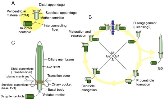
Centrosomes, cilia, and the cell cycle. (A) The centrosome is composed of mother and daughter centrioles, and a protein matrix called pericentriolar material. The mother centriole has distal and subdistal appendages. Interconnecting fibers connect the two centrioles. (B) New centrioles (procentrioles) assemble in S phase and continue to elongate in G2. The two paired centrioles separate, and the original (oldest) daughter centriole acquires appendage proteins in late G2/early M, although these appendages are not visible at this stage (appendages are depicted as dotted lines). After mitosis, the paired centrioles become disengaged. In G0, the mother centriole migrates near the plasma membrane to become a basal body, and the primary cilium is formed. (C) The basal body localizes near the plasma membrane and nucleates a primary cilium. The mother centriole converts to the basal body, and structures that include the transition fibers/distal appendage, basal foot/subdistal appendage, and striated rootlet are observed. The transition fibers tether the basal body to the plasma membrane in the transition zone, in which triplet microtubules of the basal body transition to doublet microtubules in the axoneme. The axoneme is surrounded by a ciliary membrane. Ciliary pockets are observed near basal bodies.
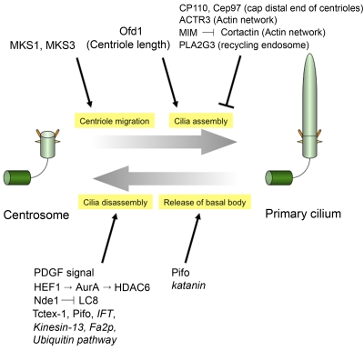
Modulating the balance between centriolar and ciliary fates. The events involved in the conversion of the centriole to the basal body and vice versa are shown. Proteins that impact these pathways are listed. Functional links to proteins in italics were shown exclusively in C. reinhardtii.
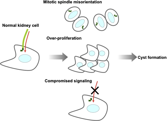
Model for cyst formation caused by loss of primary cilia in kidney cells. In normal kidney cells, mother centrioles function as basal bodies upon which primary cilia are assembled. Several signaling pathways are mediated through primary cilia (red arrows). Loss of cilia may promote defects in centrosome positioning, resulting in mitotic spindle misorientation and aberrant cell division; release of basal bodies, permitting increased proliferation; and defects in signaling pathways orchestrated by primary cilia. These abnormalities could lead to cyst formation in kidney cells.
References
Publication types
MeSH terms
LinkOut - more resources
Full Text Sources
Other Literature Sources
