. 2011 May 2;193(3):551-64.
doi: 10.1083/jcb.201011051.

WNT-3A modulates articular chondrocyte phenotype by activating both canonical and noncanonical pathways

Affiliations

WNT-3A modulates articular chondrocyte phenotype by activating both canonical and noncanonical pathways

Giovanna Nalesso et al. J Cell Biol. .

Abstract

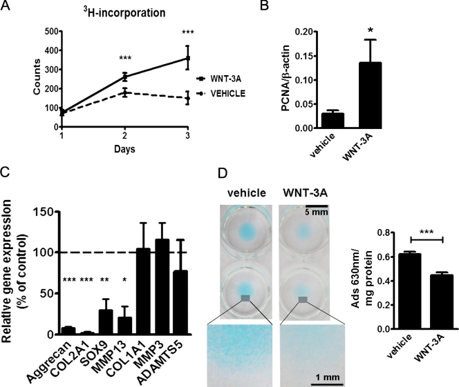
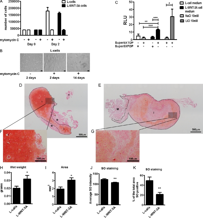
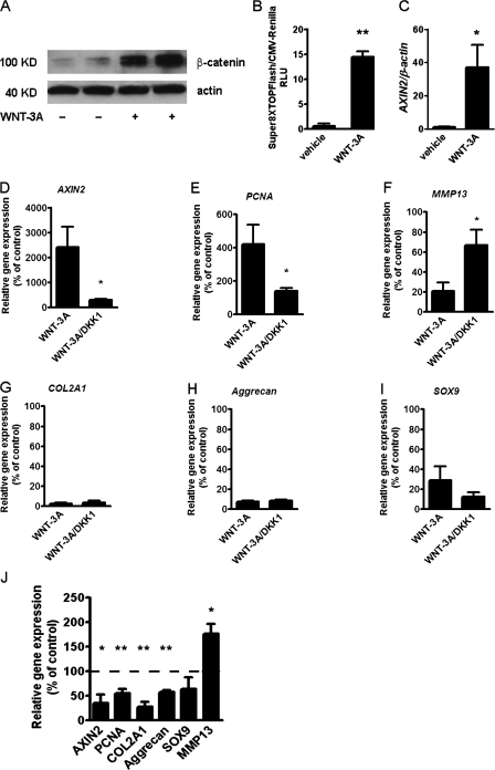
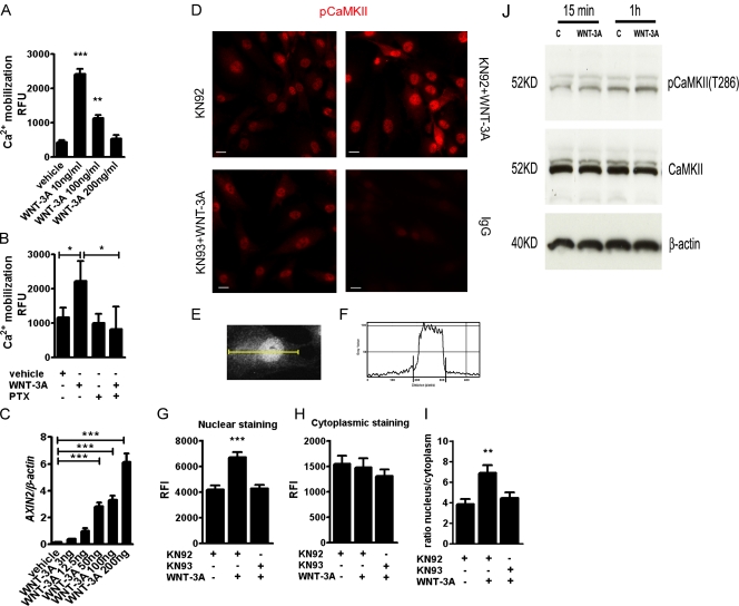
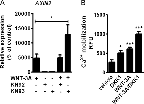
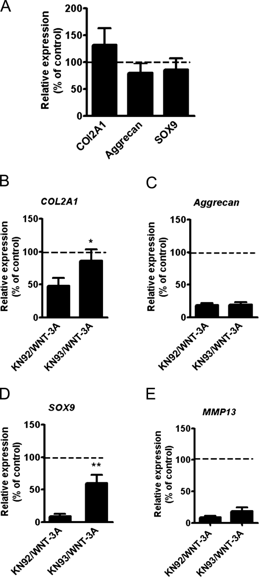
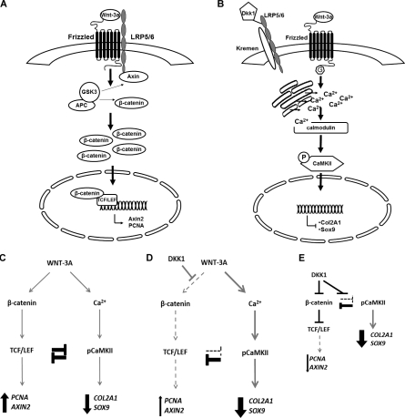
Activation and disruption of Wnt/β-catenin signaling both result in cartilage breakdown via unknown mechanisms. Here we show that both WNT-3A and the Wnt inhibitor DKK1 induced de-differentiation of human articular chondrocytes through simultaneous activation of β-catenin-dependent and independent responses. WNT-3A activates both the β-catenin-dependent canonical pathway and the Ca(2+)/CaMKII noncanonical pathways, with distinct transcriptional targets. WNT-3A promotes cell proliferation and loss of expression of the chondrocyte markers COL2A1, Aggrecan, and SOX9; however, proliferation and AXIN2 up-regulation are downstream of the canonical pathway and are rescued by DKK1, whereas the loss of differentiation markers is CaMKII dependent. Finally, we showed that in chondrocytes, the Ca(2+)/CaMKII-dependent and β-catenin-dependent pathways are reciprocally inhibitory, thereby explaining why DKK1 can induce loss of differentiation through de-repression of the CaMKII pathway. We propose a novel model in which a single WNT can simultaneously activate different pathways with distinct and independent outcomes and with reciprocal regulation. This offers an opportunity for selective pharmacological targeting.


Figures

Figure 1.
Figure 1.
WNT-3A induces chondrocyte proliferation and de-differentiation in vitro. (A and B) 100 ng/ml WNT-3A stimulated proliferation of primary chondrocytes in vitro as evaluated by an [H3]thymidine incorporation assay in porcine articular chondrocytes (A; n = 6) and by up-regulation of the proliferation marker PCNA in primary AHACs by quantitative PCR (Q-PCR) (B; n = 4). (C and D) WNT-3A–induced down-regulation of the differentiation markers COL2A1, Aggrecan, SOX9, and MMP13, as evaluated by Q-PCR (C; n = 6), and reduced the accumulation of highly sulphated GAG in primary AHACs (D; by Alcian blue staining and spectrophotometric quantification; n = 4). Gene expression values are reported as a percentage of up- or down-regulation compared with control (100%, broken line in the graph). Statistical analysis was performed with an unpaired t test. Error bars indicate mean ± SEM. *, P < 0.05; **, P < 0.005; ***, P < 0.0005.
Figure 2.
Figure 2.
WNT-3A induces proliferation and de-differentiation of articular chondrocytes in vivo. (A and B) WNT-3A induced chondrocyte de-differentiation in vivo in an ectopic cartilage formation assay. Isolated porcine articular chondrocytes were co-injected intramuscularly in nude mice together with either L cells stably expressing WNT-3A (L-WNT3A) or control L cells (L cells; 12 injections per condition). Before injection, L cells and L-WNT-3A cells were growth-arrested by mitomycin C treatment. To confirm growth arrest, 2 d after treatment, a sample of cells with or without mitomycin C treatment was detached and counted (A; n = 4). Mitomycin C–treated cells stopped proliferating. One additional well was kept in culture for 14 d. The cells persisted but never reached confluency (B). Bar, 10 µm. (C) To confirm that mitomycin C growth-arrested L-WNT-3A cells still produced biologically active WNT-3A, the conditioned medium (from the last change of medium) obtained 14 d after mitomycin C treatment was tested for the ability to activate the SUPER8XTOPFlash reporter assay in porcine articular chondrocytes. Indeed, conditioned medium from L-WNT-3A cells, but not from control cells, activated the SUPER8XTOPFlash reporter. The mutagenized SUPER8XFOPFlash vector was used to control for specificity (n = 4). (D–K) SO staining of cartilage implants obtained by the co-injection of porcine articular cartilage with L cells (D) or L-WNT-3A cells (E) in nude mice. Cartilage implants obtained from porcine articular chondrocytes co-injected with L-WNT-3A cells are on average larger (H and I) and less differentiated (J and K), as shown by a decreased SO staining in comparison with controls. Grayscale digital images from SO staining were used to calculate and compare the mean intensity of the staining and, after thresholding, the percentage of the total implant area that was positive for SO (n = 12 per condition). M, muscle; I, implant; RLU, relative luminescence unit. An unpaired t test was used for statistical evaluation. Error bars indicate mean ± SEM. *, P < 0.05; **, P < 0.005; ***, P < 0.0005.
Figure 3.
Figure 3.
Inhibition of Wnt canonical pathway with DKK1 does not rescue the loss of the chondrocyte phenotype. (A–C) WNT-3A activates the Wnt canonical pathway in AHACs. (A) Western blotting for β-catenin in the cytoplasmic fraction of AHACs treated with 100 ng/ml WNT-3A or vehicle control for 24 h. (B) WNT-3A activated the SUPER8TOPFlash reporter assay in P0 AHACs after 24 h of stimulation (n = 3). (C) WNT-3A up-regulated the expression of the endogenous Wnt canonical target gene AXIN2 in AHACs as evaluated by Q-PCR. Values were normalized for the housekeeping gene β-actin. (D–I) Primary AHACs were treated for 24 h with 100 ng/ml of recombinant WNT-3A or DKK1, alone or in combination, and were subjected to gene expression analysis by PCR. Blockade of the Wnt canonical pathway by DKK1 rescued the modulation of AXIN2, PCNA, and MMP13 mRNA (D–F) but not the down-regulation of COL2A1, Aggrecan, and SOX9 (G–I). (J) In the absence of exogenous WNT-3A, treatment with 100 ng/ml DKK1 resulted, as expected, in down-regulation of the mRNA levels of AXIN2 and PCNA, but also in a paradoxical down-regulation of COL2A1 and Aggrecan similar to that induced by WNT-3A. Q-PCR data, n = 6. Gene expression values are reported as a percentage of up- or down-regulation compared with control (100% is indicated by the broken line in the graph). Data were analyzed with an unpaired t test. Error bars indicate mean ± SEM. *, P < 0.05; **, P < 0.005.
Figure 4.
Figure 4.
WNT-3A promotes intracellular Ca2+-mediated phosphorylation of CaMKII and its nuclear translocation. (A) WNT-3A caused calcium mobilization in AHACs, particularly at low doses. Primary AHACs were treated with different amounts of recombinant WNT-3A or vehicle control for 5 min and then subjected to fluorometric determination of calcium mobilization (n = 9). (B) WNT3A-induced calcium mobilization assay in primary AHACs is G protein dependent. AHACs were preincubated overnight at 37°C with 1 µg/ml of the G protein inhibitor PTX and then treated for 5 min with 100 ng/ml of WNT-3A or vehicle control. PTX treatment blocked calcium mobilization induced by WNT-3A (n = 6). (C) Q-PCR for AXIN2 mRNA in primary chondrocytes exposed for 24 h to different doses of recombinant WNT-3A. WNT-3A induced dose-dependent up-regulation of AXIN2 in primary AHACs (n = 3). (D) WNT-3A induced nuclear accumulation of the phosphorylated form of CaMKII. Immunofluorescence staining for pCaMKII in P0 AHACs stimulated for 24 h with WNT-3A in combination with either the CaMKII inhibitor KN93 or its inactive analogue KN92 (both 10 µM). Bar, 20 µm. (E–I). Quantification of the pCaMKII fluorescence intensity in the nuclear and in the cytoplasmic fractions of stimulated AHACs. The pixel intensity profile was plotted over a linear section of a whole cell (E and F). The nuclear and cytoplasmic fluorescence intensity was then calculated as the area under the curve (G–I). (J) WNT3A promotes phosphorylation of CaMKII in T286 at 15 min and 1 h from WNT-3A stimulation. RFU, relative fluorescence unit; RFI, relative fluorescence intensity. Data were analyzed with an unpaired t test except in C, where a Kruskal Wallis with a Dunns post-test was used. Error bars indicate mean ± SEM. *, P < 0.05; **, P < 0.005; ***, P < 0.0005.
Figure 5.
Figure 5.
Wnt canonical and Wnt-CaMKII noncanonical pathways are reciprocally inhibitory in AHACs. (A) Q-PCR for AXIN2 of primary AHACs treated with 100 ng/ml WNT-3A in the presence of the CaMKII inhibitor KN93 or its inactive analogue KN92 (10 µM). (B) DKK1 promoted intracellular calcium accumulation in P0 AHACs. The inhibition of the canonical pathway with DKK1 enhanced intracellular calcium accumulation induced by WNT-3A. In all the experiments, n = 4. Data in A were analyzed with ANOVA; data in B were analyzed with an unpaired t test. Error bars indicate mean ± SEM. *, P < 0.05; ***, P < 0.0005.
Figure 6.
Figure 6.
Inhibition of the Ca2+/CaMKII pathway rescued the loss of COL2A1 and SOX9 mRNA expression in AHACs. (A) Blockade of CaMKII with KN93 did not alter the basal levels of Aggrecan, COL2A1, and SOX9. Primary AHACs were treated for 24 h with 10 µM KN93 or its inactive analogue KN92. (B–E) CaMKII blockade rescues WNT-3A–induced down-regulation of COL2A1 and SOX9 mRNA. Primary AHACs were cultured for 24 h in the presence of WNT-3A or vehicle control, and either the CaMKII inhibitor KN93 or the inactive control KN92. Gene expression was evaluated by Q-PCR. The values were normalized for β-actin and expressed as a percentage of the KN92-treated group (100% is indicated by the broken line in the graphs; n = 6). Statistical analysis was performed with an unpaired t test. Error bars indicate mean ± SEM. *, P < 0.05; **, P < 0.005.
Figure 7.
Figure 7.
Simultaneous activation and reciprocal inhibition of β-catenin– and CaMKII–dependent pathways in AHACs. (A and B) In AHACs, WNT-3A up-regulates AXIN2 expression and proliferation through the Wnt canonical pathway (A), but induces loss of differentiation via a G protein–mediated Ca2+/CaMKII–dependent pathway (B). (C–E) These two pathways are reciprocally inhibitory and in equilibrium. Therefore, either exogenous WNT-3A (C) or blockade of the canonical pathway with DKK1 (D–E) will both result in loss of chondrocyte phenotype.

Comment in

References

    1. Akiyama H., Lyons J.P., Mori-Akiyama Y., Yang X., Zhang R., Zhang Z., Deng J.M., Taketo M.M., Nakamura T., Behringer R.R., et al. 2004. Interactions between Sox9 and beta-catenin control chondrocyte differentiation. Genes Dev. 18:1072–1087 10.1101/gad.1171104 - DOI - PMC - PubMed
    1. Berridge M.J., Irvine R.F. 1989. Inositol phosphates and cell signalling. Nature. 341:197–205 10.1038/341197a0 - DOI - PubMed
    1. Chen M., Zhu M., Awad H., Li T.F., Sheu T.J., Boyce B.F., Chen D., O’Keefe R.J. 2008. Inhibition of beta-catenin signaling causes defects in postnatal cartilage development. J. Cell Sci. 121:1455–1465 10.1242/jcs.020362 - DOI - PMC - PubMed
    1. Church V., Nohno T., Linker C., Marcelle C., Francis-West P. 2002. Wnt regulation of chondrocyte differentiation. J. Cell Sci. 115:4809–4818 10.1242/jcs.00152 - DOI - PubMed
    1. Cipolletta E., Monaco S., Maione A.S., Vitiello L., Campiglia P., Pastore L., Franchini C., Novellino E., Limongelli V., Bayer K.U., et al. 2010. Calmodulin-dependent kinase II mediates vascular smooth muscle cell proliferation and is potentiated by extracellular signal regulated kinase. Endocrinology. 151:2747–2759 10.1210/en.2009-1248 - DOI - PMC - PubMed

Publication types