. 2011 May 17;108(20):8287-92.
doi: 10.1073/pnas.1105254108. Epub 2011 May 2.

Regulation of angiogenesis and choroidal neovascularization by members of microRNA-23~27~24 clusters

Affiliations

Regulation of angiogenesis and choroidal neovascularization by members of microRNA-23~27~24 clusters

Qinbo Zhou et al. Proc Natl Acad Sci U S A. .

Abstract

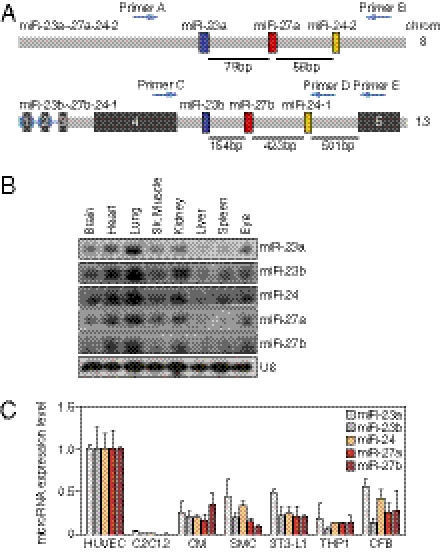
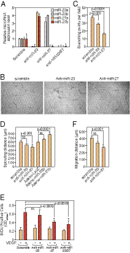
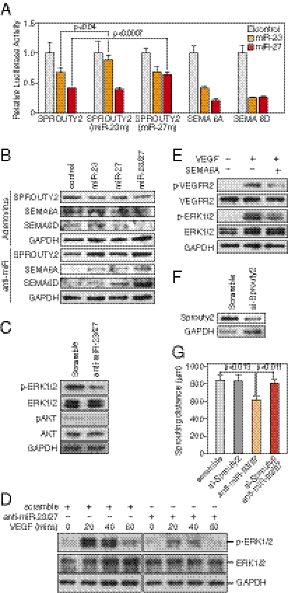
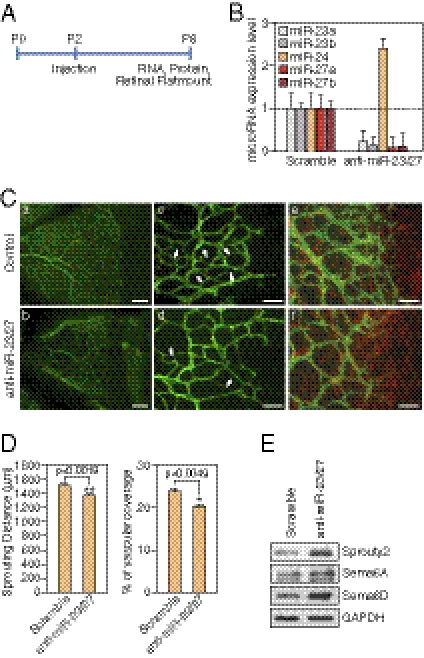
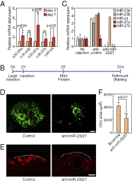
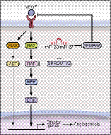
MicroRNAs (miRNAs) modulate complex physiological and pathological processes by repressing expression of multiple components of cellular regulatory networks. Here we demonstrate that miRNAs encoded by the miR-23∼27∼24 gene clusters are enriched in endothelial cells and highly vascularized tissues. Inhibition of miR-23 and miR-27 function by locked nucleic acid-modified anti-miRNAs represses angiogenesis in vitro and postnatal retinal vascular development in vivo. Moreover, miR-23 and miR-27 are required for pathological angiogenesis in a laser-induced choroidal neovascularization mouse model. MiR-23 and miR-27 enhance angiogenesis by promoting angiogenic signaling through targeting Sprouty2 and Sema6A proteins, which exert antiangiogenic activity. Manipulating miR-23/27 levels may have important therapeutic implications in neovascular age-related macular degeneration and other vascular disorders.


Conflict of interest statement

The authors declare no conflict of interest.

Figures

Fig. 1.
Fig. 1.
Gene structure and expression pattern of miR-23∼27∼24 clusters. (A) Structure of mouse miR-23∼27∼24 clusters. Chromosome locations of miR-23a∼27a∼24–2 and miR-23b∼27b∼24–1 are shown. Pre-miR23a/b, pre-miR-27a/b, and pre-miR-24 are shown as colored boxes. The exons of the miR-23b∼27b∼24–1 host gene are indicated as black boxes. (B) Expression of miR-23∼27∼24 cluster members in different tissues as detected by Northern blot using starfire miRNA probes. U6 served as a loading control. (C) Expression of miR-23∼27∼24 cluster members in different cell types relative to that in HUVECs, as detected using LNA-modified miRNA PCR primers (Exiqon). C2C12, mouse myoblast cell line; CM, mouse cardiomyocyte; SMC, mouse smooth muscle cell line; 3T3-L1, mouse adipocyte progenitor cell line; TH1, human monocyte cell line; CFB, rat cardiac fibroblast.
Fig. 2.
Fig. 2.
Regulation of angiogenesis by miR-23 and miR-27 in vitro and ex vivo. (A) Specific silencing of miR-23 and miR-27 in HUVECs by LNA-modified anti-miR shown by real-time PCR with LNA-modified miRNA primers. (B) Representative pictures of in vitro Matrigel assays after silencing of miR-23 or miR-27 in HUVECs. (C) Quantification of branching points in the in vitro Matrigel assays. Six independent samples were quantified in each group. P values are shown. (D) Quantification of ex vivo aortic ring assays 5 d after miR-23/27 knockdown or overexpression by adenovirus. Sprouting distance was measured from the average number of six aortic rings in each group. P values are indicated. ns, not significant. (E) Quantification of VEGF-induced HUVEC proliferation indicated by BrDU incorporation after miR-23 and/or miR-27 LNA-anti-miR transfection. P values are indicated. ns, not significant. (F) Quantification of VEGF-induced HUVEC migration distance (μm) in scratch wound assay after miR-23 or miR-27 LNA-anti-miR transfection. P values are indicated. ns, not significant.
Fig. 3.
Fig. 3.
Regulation of angiogenic signaling by miR-23 and miR-27. (A) miRs-23/27 target the SPROUTY2, SEMA6A, and SEMA6D 3′UTRs as shown by luciferase assays. SPROUTY2 (miR-23m) and SPROUTY2 (miR-27m) indicate SPROUTY2 UTR with mutations in miR-23 and miR-27 targeting sites, respectively. Pre-miR miRNA precursors (ABI) used in the transfections are indicated. The P values are shown. (B) Regulation of miRs-23/27 target proteins SPROUTY2, SEMA6A, and SEMA6D by miR-23 and miR-27, as detected by Western blot. LNA-anti-miR transfection or adenovirus infection was performed as indicated. (C) Repression of ERK1/2 and AKT phosphorylation by miRs-23/27 knockdown in HUVECs as shown by Western blot analyses. GAPDH served as a loading control. (D) Repression of ERK1/2 phosphorylation in response to VEGF by miRs-23/27 knockdown as shown by Western blot. LNA-anti-miR transfection and the time points of VEGF treatment are shown as indicated. (E). Repression of VEGFR2 and ERK1/2 phosphorylation in response to VEGF by recombinant SEMA6A protein as detected by Western blot. GAPDH served as a loading control. (F) Knockdown of Sprouty2 by siRNA in cultured aortas shown by Western blot. GAPDH served as a loading control. (G) Quantification of ex vivo aortic ring assays 6 d after miR-23/27 anti-miR and/or Sprouty2 siRNA transfection. Six independent aortic rings were quantified in each group. P values are indicated.
Fig. 4.
Fig. 4.
Regulation of retinal vascular development by miR-23 and miR-27. (A) Experimental setup for retinal injections. LNA-anti-miRs were injected at P2 and retinal samples are collected at P6 for RNA, protein analyses, and flatmount staining. (B) Detection of miR-23∼27∼24 family members in the retina after LNA anti-miR treatment by real-time PCR with LNA-modified miRNA primers. (C) Vasculature of the retina at P6 after LNA anti-miR treatment at P2, as visualized by ICAM-2 (green) and GFAP (red) staining in flat mount preparation. Anti-scramble (Upper) and anti-miR-23/27 (Lower). Scale bar (Left): 100 μm. Scale bar (Middle and Right): 50 μm. Arrows point to the new retinal vascular sprouts. (D) Quantification of sprouting distance (μm) and vascular coverage of the retinal vasculature from 12 miR-23/27 anti-miR treated retinas compared with 12 scramble controls. P values are indicated. (E) Western blot analyses showing the up-regulation of miRs-23/27 target proteins Sprouty2, Sema6A, and Sema6D upon miRs-23/27 knockdown in P6 retinas.
Fig. 5.
Fig. 5.
Repression of laser-induced CNV by LNA-anti-miR-23/27. (A) Up-regulation miR-23∼27∼24 cluster members 7 d after laser injury in the eye as shown by real-time PCR using LNA-miRNA primers. P values are indicated. (B) Experimental setup for laser injury and retinal injections. Time points of laser injury, anti-miR injection, and sample isolation are denoted below the line. (C) Silencing of miR-23 and miR-27 in the retina/choroid/sclera by LNA-anti-miR injection shown by real-time PCR with LNA-miRNA primers. (D) Representative confocal images of ICAM-2 staining showing repression of CNV by LNA-anti-miR-23/27 compared with a scramble control. Scale bar: 50 μm. (E) ICAM-2 immunostaining of CNV lesion sections showing reduced neovascularization by LNA-anti-miR-23/27. Dashed lines: borders the CNV lesion. Scale bar: 50 μm. (F) Quantification of CNV area (μm2). P value is from the measurements of 36 control-injected retinas and 32 LNA-anti–miR-23/27 injected retinas.
Fig. 6.
Fig. 6.
A model for miRs-23/27 function in angiogenesis. VEGF binds to its receptors and activates MAP and PI3K-AKT kinase signaling pathways in ECs, which in turn stimulates the transcription of genes involved in angiogenesis. miRs-23/27 from the miR-23∼27∼24 families promote angiogenesis by repressing their target proteins SPROUTY2 and SEMA6A, which negatively regulate Ras/MAPK signaling and VEGFR2 mediated signaling, respectively. Loss of miR-23/27 function diminishes MAPK and VEGFR2 signaling in response to VEGF, and thereby represses angiogenesis.

References

    1. Distler JH, et al. Angiogenic and angiostatic factors in the molecular control of angiogenesis. Q J Nucl Med. 2003;47:149–161. - PubMed
    1. Friedman E. Update of the vascular model of AMD. Br J Ophthalmol. 2004;88:161–163. - PMC - PubMed
    1. Jager RD, Mieler WF, Miller JW. Age-related macular degeneration. N Engl J Med. 2008;358:2606–2617. - PubMed
    1. Grisanti S, Tatar O. The role of vascular endothelial growth factor and other endogenous interplayers in age-related macular degeneration. Prog Retin Eye Res. 2008;27:372–390. - PubMed
    1. Brown DM, et al. ANCHOR Study Group. Ranibizumab versus verteporfin for neovascular age-related macular degeneration. N Engl J Med. 2006;355:1432–1444. - PubMed

Publication types