. 2011 May 17;108(20):8467-72.
doi: 10.1073/pnas.1016650108. Epub 2011 May 2.

Astroglial networks scale synaptic activity and plasticity

Affiliations

Astroglial networks scale synaptic activity and plasticity

Ulrike Pannasch et al. Proc Natl Acad Sci U S A. .

Abstract

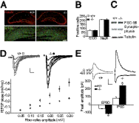
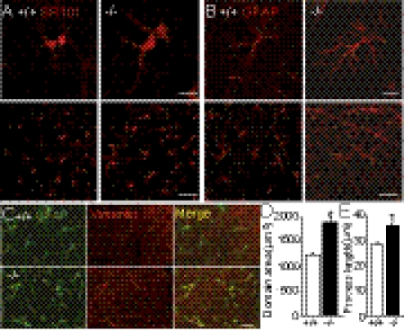
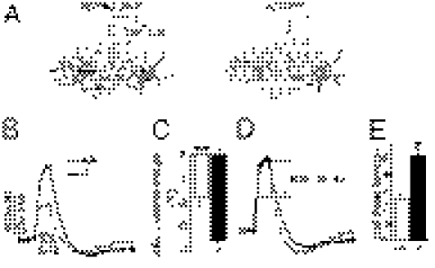
Astrocytes dynamically interact with neurons to regulate synaptic transmission. Although the gap junction proteins connexin 30 (Cx30) and connexin 43 (Cx43) mediate the extensive network organization of astrocytes, their role in synaptic physiology is unknown. Here we show, by inactivating Cx30 and Cx43 genes, that astroglial networks tone down hippocampal synaptic transmission in CA1 pyramidal neurons. Gap junctional networking facilitates extracellular glutamate and potassium removal during synaptic activity through modulation of astroglial clearance rate and extracellular space volume. This regulation limits neuronal excitability, release probability, and insertion of postsynaptic AMPA receptors, silencing synapses. By controlling synaptic strength, connexins play an important role in synaptic plasticity. Altogether, these results establish connexins as critical proteins for extracellular homeostasis, important for the formation of functional synapses.


Conflict of interest statement

The authors declare no conflict of interest.

Figures

Fig. 1.
Fig. 1.
Synaptic transmission of CA1 pyramidal cells is increased in Cx30−/−Cx43−/− mice. (A) Normal hippocampal morphology was revealed in Cx30−/−Cx43−/− hippocampal slices by immunostaining with the neuronal marker NeuN and the astrocyte marker S100. (Scale bar, 200 μm.) (B) Quantification of NeuN (Cx30−/−Cx43−/−, n = 7; WT, n = 7) and S100 positive cells (Cx30−/−Cx43−/−, n = 8; WT, n = 8,) revealed no difference in cell numbers. (C) Levels of pre- and postsynaptic markers, synaptophysin and PSD-95, respectively, are similar in Cx30−/−Cx43−/− and wild-type mice. Similar tubulin (Tub) levels confirm equivalent loading. (D) Basal excitatory transmission is enhanced in Cx30−/−Cx43−/− hippocampal slices (P < 0.05, n = 20), as assessed by input–output curves. As illustrated in the sample traces and the graph below, for each input (fiber volley, see arrow), the output (fEPSP) is increased compared with that in wild-type mice (n = 23). (Scale bar, 0.2 mV, 10 ms.) (E) Inhibitory transmission is also enhanced in CA1 pyramidal cells from Cx30−/−Cx43−/− mice (n = 5), as revealed by recordings of EPSCs (at −60 mV) and IPSCs (at 0 mV, P < 0.0001) in the same pyramidal cell (WT, n = 5). (Scale bar, 50 pA, 50 ms.)
Fig. 2.
Fig. 2.
Pre- and postsynaptic properties are altered in pyramidal cells from Cx30−/−Cx43−/− mice. (A) Release probability is increased in Cx30−/−Cx43−/− mice, as indicated by decreased paired pulse facilitation (P < 0.05, Cx30−/−Cx43−/−, n = 15; WT, n = 18). Representative EPSC traces are shown above the bar graph. (Scale bar, 10 pA, 25 ms.) (B) AMPAR activity is enhanced in Cx30−/−Cx43−/− mice, as shown by increased AMPA-NMDA EPSC ratios measured in pyramidal cells (n = 17) compared with wild-type mice (n = 21, P < 0.001). (Scale bar, 5 pA, 20 ms.) (C) Local application of AMPA (10 μM + 0.5 μM tetrodotoxin + 100 μM cyclothiazide) induced bigger responses in CA1 pyramidal cells from Cx30−/−Cx43−/− mice (P < 0.05, n = 11), compared with wild type (n = 20). (Scale bar, 100 pA, 20 s.)
Fig. 3.
Fig. 3.
Astroglial Cx43 and -30 deficiency unsilences hippocampal CA1 synapses, occludes long-term potentiation, and enhances long-term depression. (A) Reduction of silent synapses at hippocampal pyramidal cells from Cx30−/−Cx43−/− mice, illustrated by 10 superimposed traces of response at −70 mV to minimal stimulation in neurons from wild-type and Cx30−/−Cx43−/− mice. (A, Upper) Mean traces illustrate the corresponding NMDA response at +40 mV, in the presence of CNQX. (Scale bar, 5 pA, 10 ms.) (B) The enhanced release probability in Cx30−/−Cx43−/− mice was reduced to wild-type level by lowering the extracellular Ca/Mg ratio (from 2.5 mN Ca/1.3 mM Mg to 2.2 mM Ca/1.6 mM Mg) during minimal stimulation. (C) Plots of EPSC amplitude versus trial number at −70 and +40 mV. (D) Minimal stimulation revealed less AMPAR response failures and increased EPSC amplitude in pyramidal cells from Cx30−/−Cx43−/− mice (P < 0.005, n = 10) compared with wild-type mice (n = 9). (E) LTP, produced by two 100-Hz tetani separated by 20 s (arrow), was nearly absent in slices from Cx30−/−Cx43−/− mice (P < 0.005, n = 9), whereas it was induced in wild-type slices (n = 9). (E, Upper) Representative traces of averaged fEPSP recordings in slices before (solid traces) and 40–50 min after tetanic stimulation of Schaffer collaterals (shaded traces) are shown. (Scale bar, 0.05 mV, 10 ms.) (F) LTD, induced by 1-Hz stimulation over 15 min, as indicated by a solid line, was increased by ∼100% in Cx30−/−Cx43−/− mice (P < 0.05, n = 5), compared with wild-type mice (n = 5). (F, Upper) Sample traces represent averaged field potentials before (solid traces) and 50–60 min after tetanization (shaded traces). (Scale bar, 0.05 mV, 10 ms.)
Fig. 4.
Fig. 4.
Enhanced extracellular glutamate and potassium levels in Cx30−/−Cx43−/− mice due to altered astroglial clearance. (A) Synaptically evoked astrocytic GLT currents are increased in Cx30−/−Cx43−/− mice (P < 0.05, n = 6), compared with wild-type mice (n = 6). Normalization to the associated excitatory synaptic transmission (fEPSP slope/fiber volley ratio) reveals similar GLT current amplitudes. (Scale bar of representative current traces, 2.5 pA, 20 ms.) (B) GLT current decay time is prolonged in Cx30−/−Cx43−/− astrocytes (P < 0.05, n = 10) compared with wild-type astrocytes (n = 9). (C) Deconvolution-derived astroglial glutamate clearance by GLTs is slower in Cx30−/−Cx43−/− mice (P < 0.05, n = 5), compared with wild-type mice (n = 5). (C, Upper) Mean glutamate clearance time courses in wild-type and Cx30−/−Cx43−/− mice, normalized and superimposed, are shown. (Scale bar, 0.2, 10 ms.) (D) Decay time of evoked AMPAR currents, recorded in the presence of cyclothiazide (100 μM), is increased in pyramidal cells from Cx30−/−Cx43−/− mice (P < 0.01, n = 5, WT n = 6). (Scale bar, 20 pA, 50 ms.) (E) d-AA (70 μM) speeds the decay kinetics (t50) of evoked NMDAR EPSCs (recorded at −70 mV in 10 μM CNQX), only in Cx30−/−Cx43−/− mice (P < 0.05, n = 5; WT n = 5). (Scale bar of representative current traces, 10 pA, 50 ms.) (F) Synaptically evoked astrocytic potassium currents are increased in Cx30−/−Cx43−/− mice (P < 0.05, n = 8; wild type mice, n = 7). Normalization to the associated excitatory synaptic transmission is shown. (Scale bar of representative current traces, 10 pA, 1 s.) (G) Extracellular potassium rise, evoked by comparable Schaffer collateral stimulation (0.1 ms, 0.1 Hz) and monitored by astroglial membrane potential depolarization (ΔVm), is increased in Cx30−/−Cx43−/− mice (P < 0.05, n = 5; wild-type mice, n = 5). (Scale bar, 0.2 mV, 1.5 s.) (H) Kinetics of similar ΔVm amplitudes (Cx30−/−Cx43−/−, 1.1 ± 0.1 mV, n = 5 vs. WT, 0.9 ± 0.1 mV, n = 5) revealed increased half width (P < 0.05) and decay time (P < 0.01) in Cx30−/−Cx43−/− astrocytes. (Scale bar, 0.2 mV, 2 s.)
Fig. 5.
Fig. 5.
Uncoupled astrocytes are hypertrophic and reactive. (A) Cx30 and -43 deficiency causes enhanced size of stratum radiatum astrocytes, as detected by sulforhodamine-101 (SR101) labeling (Cx30−/−Cx43−/−, n = 3; WT, n = 3). (Scale bars, 10 μm and 30 μm.) (B, D, and E) Increased GFAP immunoreactivity (n = 3, B) and enlarged domain areas of Cx30−/−Cx43−/− astrocytes (P < 0.0001, n = 31; WT, n = 33, D), due to process elongation (P < 0.0001, Cx30−/−Cx43−/−, n = 54; WT, n = 54, E). (C) Astrocytes from Cx30−/−Cx43−/− mice (n = 3) are reactive, as shown by vimentin immunoreactivity (n = 3). (Scale bar, 30 μm.)
Fig. 6.
Fig. 6.
Enhanced extracellular space volume reduction during neuronal activity in Cx30−/−Cx43−/− mice. (A) Scheme illustrating the increase in extracellular concentration of TMA+ ions during neuronal activity due to cell swelling. (B) Representative traces of the time-dependent concentration change of the extracellular marker tetramethyammonium (TMA+) during 10 Hz/10 s Schaffer collateral stimulation, recorded in the stratum radiatum of the CA1 region. (C) Calculated relative changes in the extracellular space (ECS) volume (Cx43−/−Cx30−/−, n = 9; WT, n = 8). (D and E) Normalized representative TMA+ responses (D) show increased half-decay time (E) (P < 0.0001, Cx30−/−Cx43−/−, n = 9; WT, n = 8).

References

    1. Perea G, Navarrete M, Araque A. Tripartite synapses: Astrocytes process and control synaptic information. Trends Neurosci. 2009;32:421–431. - PubMed
    1. Giaume C, Koulakoff A, Roux L, Holcman D, Rouach N. Astroglial networks: A step further in neuroglial and gliovascular interactions. Nat Rev Neurosci. 2010;11:87–99. - PubMed
    1. Theis M, et al. Accelerated hippocampal spreading depression and enhanced locomotory activity in mice with astrocyte-directed inactivation of connexin43. J Neurosci. 2003;23:766–776. - PMC - PubMed
    1. Lutz SE, et al. Deletion of astrocyte connexins 43 and 30 leads to a dysmyelinating phenotype and hippocampal CA1 vacuolation. J Neurosci. 2009;29:7743–7752. - PMC - PubMed
    1. Rouach N, Koulakoff A, Abudara V, Willecke K, Giaume C. Astroglial metabolic networks sustain hippocampal synaptic transmission. Science. 2008;322:1551–1555. - PubMed

Publication types

LinkOut - more resources