. 2011 Jun;121(6):2436-46.
doi: 10.1172/JCI44796. Epub 2011 May 2.

A dual role for the immune response in a mouse model of inflammation-associated lung cancer

Affiliations

A dual role for the immune response in a mouse model of inflammation-associated lung cancer

Michael Dougan et al. J Clin Invest. 2011 Jun.

Abstract

Lung cancer is the leading cause of cancer death worldwide. Both principal factors known to cause lung cancer, cigarette smoke and asbestos, induce pulmonary inflammation, and pulmonary inflammation has recently been implicated in several murine models of lung cancer. To further investigate the role of inflammation in the development of lung cancer, we generated mice with combined loss of IFN-γ and the β-common cytokines GM-CSF and IL-3. These immunodeficient mice develop chronic pulmonary inflammation and lung tumors at a high frequency. Examination of the relationship between these tumors and their inflammatory microenvironment revealed a dual role for the immune system in tumor development. The inflammatory cytokine IL-6 promoted optimal tumor growth, yet wild-type mice rejected transplanted tumors through the induction of adaptive immunity. These findings suggest a model whereby cytokine deficiency leads to oncogenic inflammation that combines with defective antitumor immunity to promote lung tumor formation, representing a unique system for studying the role of the immune system in lung tumor development.


Figures

Figure 1
Figure 1. BALB/c TKO mice develop lung cancer.
Survival curves compare BALB/c WT (n = 14), BALB/c TKO (n = 37), and BL6 TKO (n = 27) mice.
Figure 2
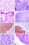
Figure 2. BALB/c TKO lung tumors are invasive pulmonary adenocarcinomas.
(AD) H&E stains on formalin-fixed tissue sections. One-year-old WT (A) and TKO (B) lung (original magnification, ×40). (C and D) TKO lung showing a dysplastic lesion (C; ×400) in a 6-month-old mouse and invasive pulmonary adenocarcinoma (D; ×20) in a 14-month-old animal. Arrows in C indicate dysplastic epithelium. SPC (E) and pERK (F) immunohistochemistry (×100) on BALB/c TKO pulmonary adenocarcinoma from 16-month-old (E) and 12-month-old (F) mice. The tumor (Tu) is indicated. (G and H) H&E staining showing invasion (G; ×100) indicated by the arrows and thoracic lymph node metastasis (Met) (H; ×20) in a 15-month-old mouse; G shows a detail from D.
Figure 3
Figure 3. BALB/c TKO lung tumors develop in the context of pulmonary and systemic inflammation.
(A) Comparison of total leukocytes recovered from BAL fluid from 3-month-old BALB/c TKO and WT mice (left panel) and analysis of lymphoid and myeloid subtypes (middle panel) using flow cytometry. Right panel: Total spleen cells from the same WT and TKO mice are presented for comparison. (B) Quantification of inflammatory cytokines present in the BAL fluid (left panel) and serum (right panel) of 3-month-old BALB/c TKO and WT mice; each point represents an individual animal. Cytokines were measured using fluorescent anti-cytokine beads; IL-6 levels in the BAL fluid were confirmed by ELISA. Results represent an analysis of samples collected from 3 independent experiments. (C and D) Quantification of serum cytokines in aged BALB/c WT and TKO mice by ELISA. (C) Points represent individual animals. (D) Serum IL-17 in 1-year-old WT (n = 6) and TKO (n = 4) mice. (E and F) Cytokine production from 1 × 10⁶ CD4+ T cells (E) stimulated for 48 hours using anti-CD3 (10 μg/ml) and anti-CD28 (2 μg/ml), or from CD11b+ macrophages (F) cultured for 18 hours. Cells were isolated from spleens of 6- to 8-week-old mice by positive selection with magnetic beads (Miltenyi Biotec). Cytokines/chemokines were measured using anti-cytokine beads. Results are combined from 2 independent experiments with a total of 6 (E) or 4 (F) mice per group. Error bars represent SEM.
Figure 4
Figure 4. Transplanted BALB/c TKO tumor cell lines elicit a vigorous host response.
(A) Analysis of cytokine secretion by BALB/c TKO lung tumor cells lines (MDAC1, MDAC8) using anti-cytokine fluorescent beads. *P < 0.05, MDAC8 compared with LL (B) Cyst associated with a transplanted BALB/c TKO lung tumor. (C) Analysis of cyst contents by flow cytometry using the indicated antibodies; numbers are an average from 2 tumor-associated cysts harvested from separate animals. (D) Quantification of immune cell expansion in the spleens of mice harboring transplanted BALB/c TKO tumors. Cell types were identified by flow cytometry. Results are combined from 3 independent experiments with 2–3 animals per group. Error bars represent SEM. *P < 0.05, **P < 0.005, MDAC8-injected compared with control (–) animals.
Figure 5
Figure 5. Knockdown of IL-6 reduces the proliferation of a BALB/c TKO lung tumor cell line.
(A) 1 × 10⁴ MDAC8 cells infected with lentiviruses encoding nontargeting shRNA (scr), or shRNA against IL-6 (KD-1 and KD-2) were cultured for 3 days in RPMI; supernatants were harvested, and total IL-6 was measured by ELISA. (B) Western blot for cytoplasmic pERK (top panels) or nuclear (nuc) STAT3 (bottom panels) using lysates from IL-6–knockdown or control (scr) MDAC8 cells. (C and D) In vitro (C) and in vivo (D) growth of MDAC8 cells expressing IL-6 shRNA compared with cells expressing nontargeting controls. (C) 1 × 10⁴ cells were cultured in RPMI. Growth was measured by CellTiter-Glo (CTG, Promega). (D) Left panel: 4 × 10⁴ cells were injected subcutaneously into BALB/c TKO mice, with 8 animals per group. Right panel: Survival of mice harboring tumors. (E) Comparison of IL-6 production from ex vivo cultured IL-6 KD-2 or control MDAC8 cell lines following growth in a secondary host (ex vivo) or after in vitro culture (baseline). Three to 4 tumors were used per group. (AE) Results are representative of at least 2 independent experiments, with 6–8 replicates per experiment. Error bars represent the SEM for the experiment shown.
Figure 6
Figure 6. Inhibition of NF-κB signaling in a BALB/c TKO lung tumor cell line leads to rapid loss of viability.
(A) Western blot of nuclear lysates from MDAC8 cells cultured for 1 hour with an IKK2 inhibitor (IKK2i), a combined IKK1-IKK2 inhibitor (IKK1/2i), or vehicle (DMSO); Data in A and B represent samples analyzed from 2 independent cultures. (B) 3 × 10⁵ MDAC8 cells were cultured for 72 hours with IKK inhibitors, vehicle (DMSO), or media (No Tx); IKK2 inhibitor I is identical to IKK2i in A, and IKK1/2 inhibitor II is identical to IKK1/2i in A; relative cell numbers were determined using CTG, with the average reading for the untreated group defined as 1. (C) Time course of cell loss following IKK inhibitor treatment; cells were treated as in B and analyzed using CTG; the average reading for the untreated group at time 0 was defined as 1. (D) Dose response to IKK1/2 inhibitor II after 72 hours; cells were treated and analyzed as in B. (E) IL-6 production from MDAC8 cells treated with IKK inhibitors as in B; IL-6 was measured by ELISA. (BE) Results are representative of 2–3 independent experiments, with 6 replicates per group. Error bars represent SEM. All IKK inhibitors were from EMD Biosciences and were used at the following concentrations unless otherwise indicated: IKK2 inhibitor I (EMD IKK-2 Inhibitor IV), 10 μM; IKK2 inhibitor II (EMD IKK-2 Inhibitor VI), 5 μM; IKK1/2 inhibitor I (BMS-345541), 40 μM; IKK1/2 inhibitor II (EMD IKK Inhibitor VII), 20 μM.
Figure 7
Figure 7. Growth of BALB/c TKO lung tumors is restricted in immunocompetent mice.
(A) Analysis of tumor-infiltrating leukocytes by flow cytometry using the indicated antibodies; numbers are an average from 4 tumors harvested in 2 independent experiments. (B and C) 4 × 10⁴ MDAC8 cells were transplanted subcutaneously into BALB/c WT or BALB/c TKO hosts. (B) Quantification of tumor size following transplantation. (C) Survival curve for mice following transplantation. (D and E) MDAC8 cells were harvested from either WT (MDAC8.WT) or TKO hosts (MDAC8.TKO). (D) 4 × 10⁴ MDAC8.WT or MDAC8.TKO cells were plated in RPMI. Proliferation was measured by CTG; linear regression analysis on log-transformed data was used to calculate doubling time. (E) 4 × 10⁴ MDAC8.WT or MDAC8.TKO cells were transplanted into WT or TKO hosts. (F and G) 4 × 10⁴ MDAC8 cells were transplanted subcutaneously into BALB/c WT or BALB/c RAG KO hosts. (F) Quantification of tumor size following transplantation. (G) Survival curve for mice following transplantation. (BG) Error bars represent SEM. Results are representative of at least 2 independent experiments with 8 to 12 mice per group.

References

    1. Jemal A, et al. Cancer statistics, 2008. CA Cancer J Clin. 2008;58(2):71–96. doi: 10.3322/CA.2007.0010. - DOI - PubMed
    1. Travis WD, Travis LB, Devesa SS. Lung cancer. Cancer. 1995. 75 (1 suppl) : 191 202 10.1002/1097-0142(19950101)75:1+<191::AID-CNCR2820751307>3.0.CO;2-Y - DOI - PubMed
    1. Doll SR. Smoking and lung cancer. Am J Respir Crit Care Med. 2000;162(1):4–6. - PubMed
    1. Kuper H, Adami HO, Boffetta P. Tobacco use, cancer causation and public health impact. J Intern Med. 2002;251(6):455–466. doi: 10.1046/j.1365-2796.2002.00993.x. - DOI - PubMed
    1. Herbst RS, Heymach JV, Lippman SM. Lung cancer. N Engl J Med. 2008;359(13):1367–1380. doi: 10.1056/NEJMra0802714. - DOI - PMC - PubMed

Publication types

MeSH terms