. 2011 Jun;121(6):2361-70.
doi: 10.1172/JCI45213. Epub 2011 May 2.

Transcription intermediary factor 1γ is a tumor suppressor in mouse and human chronic myelomonocytic leukemia

Affiliations

Transcription intermediary factor 1γ is a tumor suppressor in mouse and human chronic myelomonocytic leukemia

Romain Aucagne et al. J Clin Invest. 2011 Jun.

Abstract

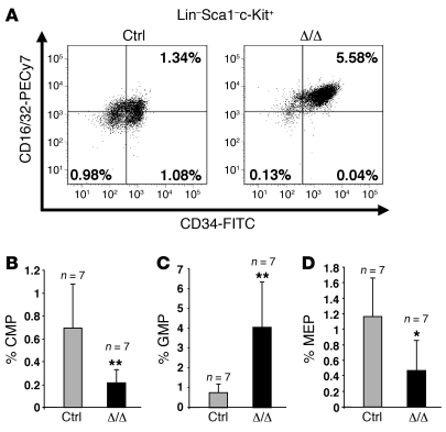
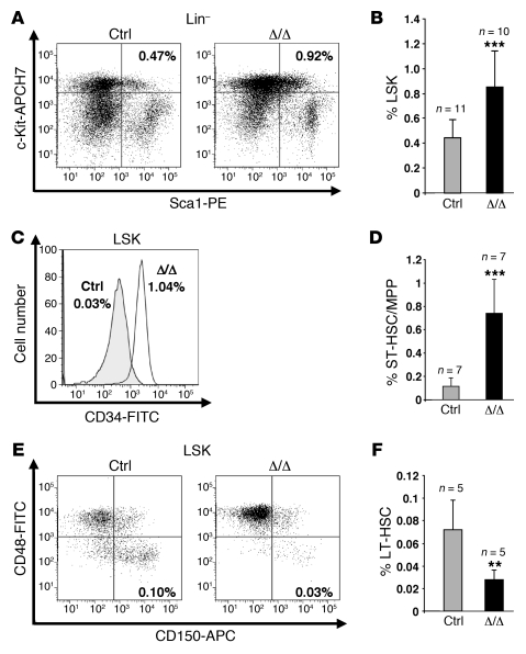
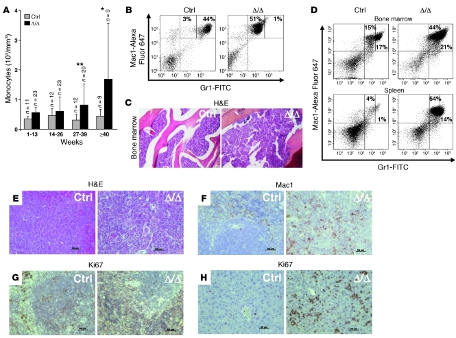
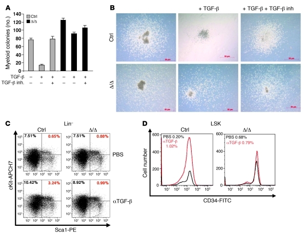
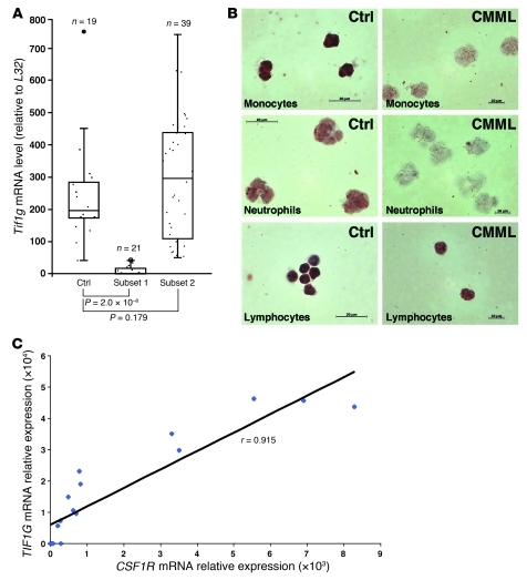
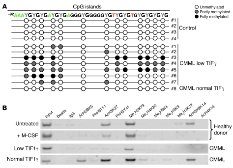
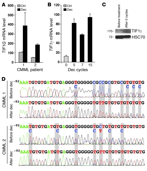
Transcription intermediary factor 1γ (TIF1γ) was suggested to play a role in erythropoiesis. However, how TIF1γ regulates the development of different blood cell lineages and whether TIF1γ is involved in human hematological malignancies remain to be determined. Here we have shown that TIF1γ was a tumor suppressor in mouse and human chronic myelomonocytic leukemia (CMML). Loss of Tif1g in mouse HSCs favored the expansion of the granulo-monocytic progenitor compartment. Furthermore, Tif1g deletion induced the age-dependent appearance of a cell-autonomous myeloproliferative disorder in mice that recapitulated essential characteristics of human CMML. TIF1γ was almost undetectable in leukemic cells of 35% of CMML patients. This downregulation was related to the hypermethylation of CpG sequences and specific histone modifications in the gene promoter. A demethylating agent restored the normal epigenetic status of the TIF1G promoter in human cells, which correlated with a reestablishment of TIF1γ expression. Together, these results demonstrate that TIF1G is an epigenetically regulated tumor suppressor gene in hematopoietic cells and suggest that changes in TIF1γ expression may be a biomarker of response to demethylating agents in CMML.


Figures

Figure 1
Figure 1. The Tif1g deletion affects hematopoietic progenitor populations in mice younger than 6 months.
(A) Representative FACS staining profiles of the progenitor populations, including CMPs (lower right panel), MEPs (lower left panel), and GMPs (upper right panel), from the respective control (Ctrl) or Tif1gΔ/Δ (Δ/Δ) mice. Calculation of the numbers and percentages of each of the populations was based on the number of living cells. The number of CMPs and MEPs decreased, whereas the number of GMPs increased. Analysis of control and Tif1gΔ/Δ mice demonstrated a decrease in CMPs (B), an increase in GMPs (C), and a decrease in MEPs (D) in Tif1gΔ/Δ mice. *P < 0.05, **P < 0.01.
Figure 2
Figure 2. The Tif1g deletion affects HSCs in mice younger than 6 months.
(A and B) Analysis of LSK cells from representative control and healthy Tif1gΔ/Δ mice demonstrated an increase in the LSK population in Tif1gΔ/Δ mice. (C and D) Analysis of ST-HSCs/MPPs from representative control and Tif1gΔ/Δ mice demonstrated an increase in ST-HSCs/MPPs in Tif1gΔ/Δ mice. (E and F) Analysis of LT-HSCs from representative control and Tif1gΔ/Δ mice demonstrated a decrease in the LT-HSC population in Tif1gΔ/Δ mice. **P < 0.01, ***P < 0.001.
Figure 3
Figure 3. Tif1gΔ/Δ mice older than 6 months develop a CMML-like myeloproliferative disease with monocytic features.
(A) Peripheral monocyte counts in control and Tif1gΔ/Δ mice. The results are shown as mean ± SD (n = 9–23 mice). (B) FACS analysis of peripheral blood cells demonstrated an increase in the monocytic (Gr1loMac1+) population in Tif1gΔ/Δ mice. (C) Representative H&E-stained histopathologic sections of bone marrow demonstrated hypercellularity in Tif1gΔ/Δ mice. (D) FACS analyses of the cell populations from bone marrow or spleen demonstrated an increase of the monocytic (Gr1loMac1+) population in Tif1gΔ/Δ mice. (E) Representative H&E-stained histopathologic sections of spleen demonstrated that the splenic organization was destroyed in Tif1gΔ/Δ mice. (F) Immunohistochemical (IHC) staining for Mac1 in paraffin-embedded spleen sections indicated an expanded monocytic population. (G and H) IHC staining for Ki67 in paraffin-embedded spleen (G) or liver (H) revealed a highly proliferative population. Original magnification, ×76. *P < 0.05, **P < 0.01.
Figure 4
Figure 4. Alteration of the TGF-β pathway in aging Tif1gΔ/Δ mice.
(A) Defective TGF-β signaling pathway responsiveness in Tif1gΔ/Δ mice. Sorted ST-HSCs/MPPs were plated on methylcellulose medium in triplicate, with or without TGF-β and TGF-β inhibitor (inh.). The number of myeloid colonies was determined at day 8. The results are shown as the mean ± SD of triplicates. (B) Representative images (original magnification, ×138) of the resulting myeloid colonies from sorted ST-HSCs/MPPs, untreated or treated with TGF-β or with TGF-β plus TGF-β inhibitor. (C and D) TGF-β–neutralizing antibody (αTGF-β) does not affect hematopoietic progenitor cell distribution in Tif1gΔ/Δ mice. (C) Analysis of LSK cells from treated or untreated representative control and Tif1gΔ/Δ mice demonstrated an increase in the LSK population in control mice treated with the antibody, which was not observed in Tif1gΔ/Δ mice. (D) Analysis of ST-HSCs/ MPPs from treated or untreated representative control and Tif1gΔ/Δ mice demonstrated an increase in MPPs in control mice treated with the antibody, which was not observed in Tif1gΔ/Δ mice.
Figure 5
Figure 5. TIF1γ is downregulated in 35% of CMML patients.
(A) RQ-PCR analysis of TIF1γ expression in the purified monocytes (CD14+ cells) of healthy donors (Ctrl) or CMML patients (subsets 1 and 2). (B) Immunocytochemistry staining for TIF1γ from the monocytes, neutrophils and lymphocytes (×340) of a representative control or CMML patient. (C) Correlation of TIF1G and CSF1R expression in monocytes of 17 individuals with CMML. The scatter plot shows TIF1G and CSF1R mRNA expression. Expression values are expressed in arbitrary units. A linear regression curve and the correlation coefficient are indicated. *P < 0.05.
Figure 6
Figure 6. TIF1G is an epigenetically regulated tumor suppressor gene in CMML.
(A) Sequencing of the bisulfite modified TIF1G promoter sequence from normal monocytes (Control) or CMML monocytes. (B) ChIP analysis of the status of histone modifications on the TIF1G promoter. Chromatin was immunoprecipitated using specific antibodies directed against histone modifications. Ac, acetylated; Me1, methylated; Me3, tri-methylated.
Figure 7
Figure 7. TIF1G is sensitive to decitabine in CMML.
(A) TIF1G expression (RQ-PCR) from the monocytes (CD14+) of two CMML patients (1 and 2) after 3 days of culture in the absence or presence of decitabine (dec). Data are the mean ± SD of the values from experiments performed in triplicate. (B) TIF1G expression (RQ-PCR) from the monocytes of one CMML patient before and during treatment with decitabine. Data are the mean ± SD of the values from experiments performed in triplicate. (C) Immunoblotting analysis for TIF1γ in human monocytes from the same patient after 5 cycles of decitabine. Equivalent loading of the lanes was controlled by anti-HSC70 antibody staining. (D) Sequencing of the bisulfite-modified TIF1G promoter sequence from the monocytes of two patients before and after 7 cycles of decitabine (CMML 1 is the same patient as in B).

References

    1. Venturini L, et al. TIF1gamma, a novel member of the transcriptional intermediary factor 1 family. Oncogene. 1999;18(5):1209–1217. doi: 10.1038/sj.onc.1202655. - DOI - PubMed
    1. Beckstead R, et al. Bonus, a Drosophila homolog of TIF1 proteins, interacts with nuclear receptors and can inhibit betaFTZ-F1-dependent transcription. Mol Cell. 2001;7(4):753–765. doi: 10.1016/S1097-2765(01)00220-9. - DOI - PMC - PubMed
    1. Friedman JR, et al. KAP-1, a novel corepressor for the highly conserved KRAB repression domain. Genes Dev. 1996;10(16):2067–2078. doi: 10.1101/gad.10.16.2067. - DOI - PubMed
    1. Khetchoumian K, et al. TIF1delta, a novel HP1-interacting member of the transcriptional intermediary factor 1 (TIF1) family expressed by elongating spermatids. J Biol Chem. 2004;279(46):48329–48341. doi: 10.1074/jbc.M404779200. - DOI - PubMed
    1. Le Douarin B, et al. The N-terminal part of TIF1, a putative mediator of the ligand-dependent activation function (AF-2) of nuclear receptors, is fused to B-raf in the oncogenic protein T18. EMBO J. 1995;14(9):2020–2033. - PMC - PubMed

Publication types

MeSH terms