

Review
doi: 10.1007/s10969-011-9110-6.
Epub 2011 May 3.
Beyond structural genomics: computational approaches for the identification of ligand binding sites in protein structures
Affiliations
- PMID: 21537951
- PMCID: PMC3127736
- DOI: 10.1007/s10969-011-9110-6
Item in Clipboard
Review
Beyond structural genomics: computational approaches for the identification of ligand binding sites in protein structures
J Struct Funct Genomics.
2011 Jul.
Abstract
Structural genomics projects have revealed structures for a large number of proteins of unknown function. Understanding the interactions between these proteins and their ligands would provide an initial step in their functional characterization. Binding site identification methods are a fast and cost-effective way to facilitate the characterization of functionally important protein regions. In this review we describe our recently developed methods for binding site identification in the context of existing methods. The advantage of energy-based approaches is emphasized, since they provide flexibility in the identification and characterization of different types of binding sites.
Figures
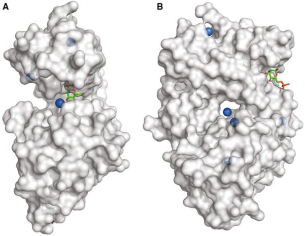
Geometric identification of binding sites. a Human alpha-Phosphomannomutase in complex with d -mannose 1-phosphate (PDB code: 2fue [48]). The top three binding sites identified by LIGSITEcsc are represented by blue spheres. The ligand binds in a deep crevice that is correctly identified as the largest pocket. b Mannose 6-phosphate receptor in complex with mannose 6-phosphate (PDB code: 1sz0 [49]). The binding site is a shallow pocket and in this case is not among the top five sites predicted by LIGSITEcsc. The blue spheres show the pockets identified by LIGSITEcsc
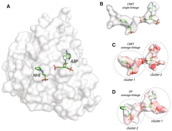
Energy-based identification of binding sites using SiteHound. a Structure of human pregnenolone sulfotransferase bound to Adenosine-3′-5′-diphosphate (A3P) and 2-[N-cyclohexylamino] ethane sulfonic acid (NHE) (PDB code: 1q1q [50]). b Identification of the ligand binding site region using the methyl (CMET) probe and single-linkage clustering. A single cluster covers the two ligands and the entrance to the ligand-binding channel. c Identification of the binding sites for NHE (cluster 1) and A3P (cluster 2) using the CMET probe and average linkage clustering. The two binding-sites are identified as separate ligands. The clusters are colored according to the local interaction energy, with red corresponding to stronger interactions. d Identification of the binding sites for A3P (cluster 1) and NHE (cluster 2) using the phosphate oxygen (OP) probe. Note the reversal of the cluster ranking, with the cluster for the phosphate-containing ligand (A3P) ranking first, and the most favorable interaction energy spots (red regions) being located around the phosphate and sulfonate groups of the ligands
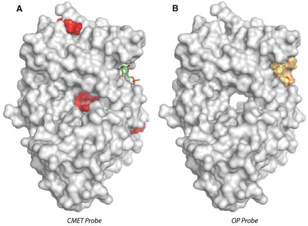
Identification of a shallow binding site using the OP probe in SiteHound. Mannose 6-phosphate receptor in complex with mannose 6-phosphate (see Fig. 1). a The top 5 clusters identified with the methyl (CMET) probe of SiteHound are shown as red surfaces. Two clusters are located on the opposite side of the structure and not visible. b Top 3 clusters identified with the phosphate oxygen (OP) probe of SiteHound are shown as orange surfaces. Two clusters are located on the opposite side of the structure and not visible
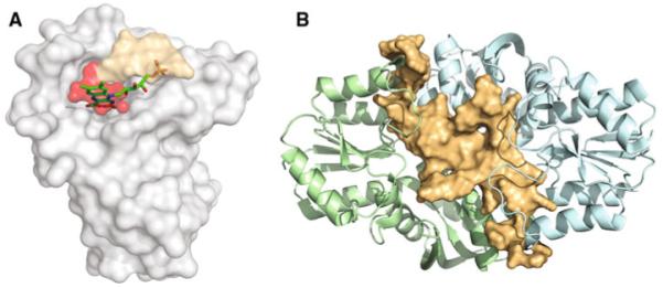
Examples of binding site identification in proteins of unknown function. a Structure of Rv2074 from Mycobacterium tuberculosis (PDB code: 2asf [51]) showing a binding site identified by the CMET probe (red) and OP probe (orange) of SiteHound. Rv2074 has been suggested as a probable pyridoxine 5′-phosphate oxidase [51]. The clusters delineate a putative binding site that occupies the same position as the Flavin mononucleotide molecule (shown) in the structurally similar Rv1155 pyridoxine 5′-phosphate oxidase [52]. b Structure of Escherichia coli protein ybgI (PDB code: 1nmo [53]) showing a large OP probe cluster identified using the single-linkage clustering algorithm in SiteHound. This large cluster would seem to be compatible with a suggested role of ybgI in DNA metabolism [53]
References
-
- Brent MM, Marmorstein R. Ankyrin for methylated lysines. Nat Struct Mol Biol. 2008;15:221–222. - PubMed
-
- Shima S, Pilak O, Vogt S, Schick M, Stagni MS, Meyer-Klaucke W, Warkentin E, Thauer RK, Ermler U. The crystal structure of [Fe]-hydrogenase reveals the geometry of the active site. Science. 2008;321:572–575. - PubMed
-
- Copley SD. Enzymes with extra talents: moonlighting functions and catalytic promiscuity. Curr Opin Chem Biol. 2003;7:265–272. - PubMed
-
- Jeffery CJ. Moonlighting proteins: old proteins learning new tricks. Trends Genet. 2003;19:415–417. - PubMed
Publication types
MeSH terms
Substances
Grants and funding
LinkOut - more resources
Full Text Sources
