. 2011 Aug;34(4):343-54.
doi: 10.1007/s13402-011-0035-7. Epub 2011 May 3.

Rapid quantification of myocardial fibrosis: a new macro-based automated analysis

Affiliations

Rapid quantification of myocardial fibrosis: a new macro-based automated analysis

Awal M Hadi et al. Cell Oncol (Dordr). 2011 Aug.

Abstract

Background: Fibrosis is associated with various cardiac pathologies and dysfunction. Current quantification methods are time-consuming and laborious. We describe a semi-automated quantification technique for myocardial fibrosis and validated this using traditional methods.

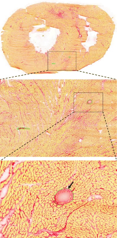
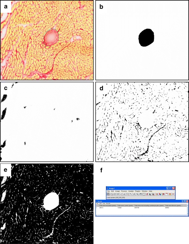
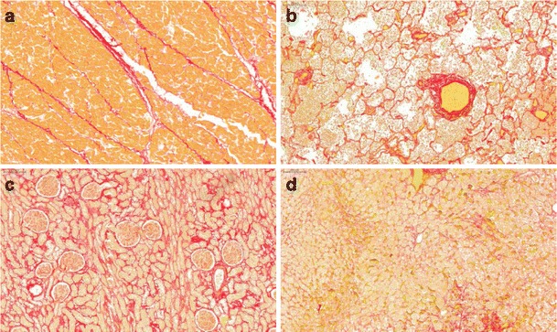
Methods: Pulmonary Hypertension (PH) was induced in adult Wistar rats by subcutaneous monocrotaline (MCT) injection(40 mg/kg). Cryosections of myocardial tissue (5 μm) of PH rats (n = 9) and controls (n = 9) were stained using Picrosirius red and scanned with a digital microscopic MIRAX slide scanner. From these sections 21 images were taken randomly of each heart. Using ImageJ software a macro for automated image analysis of the amount of fibrosis was developed. For comparison, fibrosis was quantified using traditional polarisation microscopy. Both methods were correlated and validated against stereology as the gold standard. Furthermore, the method was tested in paraffin-embedded human tissues.

Results: Automated analysis showed a significant increase of fibrosis in PH hearts vs. control. Automated analysis correlated with traditional polarisation and stereology analysis (r(2) = 0.92 and r(2) = 0.95 respectively). In human heart, lungs, kidney, and liver, a similar correlation with stereology (r(2) = 0.91) was observed. Time required for automated analysis was 22% and 33% of the time needed for stereology and polarisation analysis respectively.

Conclusion: Automated quantification of fibrosis is feasible, objective, and time-efficient.


Figures

Fig. 1
Fig. 1
The Mirax scan was used to scan entire sections of Picrosirius red stained myocardial. The Mirax Viewer software allows the user to navigate through the section and zoom until the desired magnification used for analyses of fibrosis. If the user wishes to exclude areas, like staining artifacts or vessels, the user can apply annotations to the image by demarcating them in (in our study) green (arrow)
Fig. 2
Fig. 2
This figure shows the different steps of the ImageJ macro in identifying different structures in a (a) Picrosiriusred stained myocardial crossection. (b) First the macro determines the area to excluded which is demarcated by the investigator. (c) Subsequently the areas not occupied by structures are analysed and determined as lumen. (d) The macro then analyses the area occupied by fibrosis and by subtracting these values, the area occupied by (e) cardiomyocytes (in this study; this image contains all tissue compartments left) is determined. At last (f) the results in pixels are presented to the user
Fig. 3
Fig. 3
This figure shows images used in the quantification of fibrosis using polarisation light microscopy. (a) Shows a typical example of a Picrosirius red stained section of myocardium. (b) Using polarisation filters, only polarised light is passed through which can be transferred into (c) a binary image, used for calculation of the percentage fibrosis per area
Fig. 4
Fig. 4
Polarisation and ImageJ analyses showed that hearts of PH rats revealed more fibrosis compared to control. Note the increase in collagen content was not limited to the RV only, but was also found in the LV and the septum of these hearts. Note also that ImageJ analyses detected more fibrosis than the method of polarization
Fig. 5
Fig. 5
We compared both analysis methods with traditional quantification of fibrosis using stereology. Both polarisation light microscopy (a) and the ImageJ macro (b) correlated strongly with stereology (r2 = 0.93, p < 0.0001, intercept at X = 0 is −0.09 and r2 = 0.95, p < 0.0001, intercept at X = 0 is 0.32, respectively). In addition, the ImageJ macro (c) correlated with polarisation microscopy as well (r2 = 0.92, p < 0.0001, intercept at X = 0 is 0.37)
Fig. 6
Fig. 6
Picrosirius red staining was also performed on human (a) myocardial tissue, (b) lung, (c) kidney and (d) liver and scanned with Mirax scan
Fig. 7
Fig. 7
The Image J macro was able to detect fibrosis in paraffin embedded sections of human tissue of heart, lung, kidney and liver in the same way as in cardiac tissue of rat and a similar correlation was found with stereology (r2 = 0.91)

Corrected and republished from

Similar articles

Cited by

References

    1. Akhavein F, St-Michel EJ, Seifert E, Rohlicek CV. Decreased left ventricular function, myocarditis, and coronary arteriolar medial thickening following monocrotaline administration in adult rats. J Appl Physiol. 2007;103:287–295. doi: 10.1152/japplphysiol.01509.2005. - DOI - PubMed
    1. Berk BC, Fujiwara K, Lehoux S. ECM remodeling in hypertensive heart disease. J Clin Invest. 2007;117:568–575. doi: 10.1172/JCI31044. - DOI - PMC - PubMed
    1. Borbely A, Papp VZ, Bronzwaer JG, Edes I, Stienen GJ, Paulus WJ. Cardiomyocyte stiffness in diastolic heart failure. Circulation. 2005;111:774–781. doi: 10.1161/01.CIR.0000155257.33485.6D. - DOI - PubMed
    1. Brilla CG, Pick R, Tan LB, Janicki JS, Weber KT. Remodeling of the rat right and left ventricles in experimental hypertension. Circ Res. 1990;67:1355–1364. - PubMed
    1. Burstein B, Nattel S. Atrial fibrosis: mechanisms and clinical relevance in atrial fibrillation. J Am Coll Cardiol. 2008;51:802–809. doi: 10.1016/j.jacc.2007.09.064. - DOI - PubMed

Publication types

MeSH terms

LinkOut - more resources