. 2011 Jul;41(7):1832-42.
doi: 10.1002/eji.201041258. Epub 2011 Jun 6.

NKG2A is a marker for acquisition of regulatory function by human CD8+ T cells activated with anti-CD3 antibody

Affiliations

NKG2A is a marker for acquisition of regulatory function by human CD8+ T cells activated with anti-CD3 antibody

Vitaly Ablamunits et al. Eur J Immunol. 2011 Jul.

Abstract

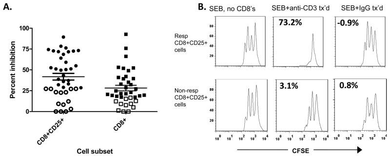
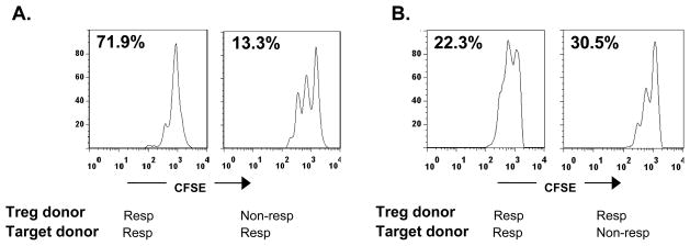
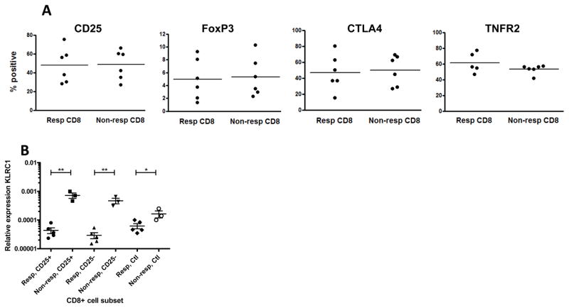
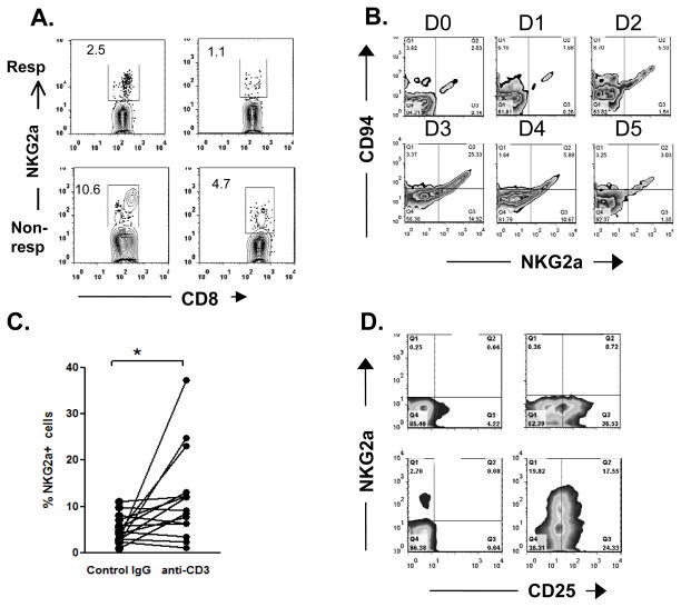
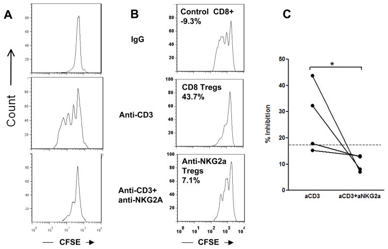
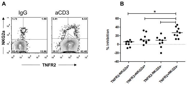
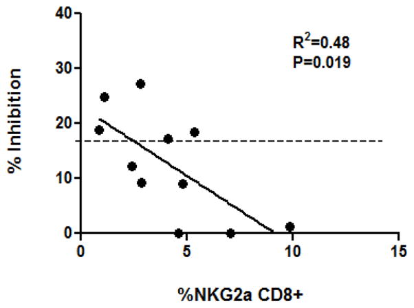
Treatment with anti-CD3 mAb modulates immune responses that cause type 1 diabetes and other diseases. CD8+ Tregs can be induced in vitro and in vivo by mAb. However, 1/3 of patients do not respond to drug therapy and in an equal proportion, anti-CD3 mAb does not induce Tregs in vitro. The acquisition of CD8+ Treg activity is a function of the CD8+ cells and not the targets in the assay. To identify markers to differentiate responses of CD8+ Tregs, we analyzed genes differentially expressed in CD8+ T cells of non-responders compared with responders, and found that an inhibitory receptor NKG2A (CD159a) was highly expressed in cells from all non-responders tested. Application of a mAb agonistic to NKG2A during in vitro CD8+ Treg induction by anti-CD3 prevented induction of CD8+ Tregs. CD8+ T cells that are TNFR2+ but NKG2A- are the most potently induced Tregs. The level of NKG2A expression on resting CD8+ T cells inversely correlated with acquisition of regulatory function when activated. We suggest that the induction of human CD8+ Tregs by anti-CD3 mAb is controlled by a negative signaling through NKG2A, and that NKG2A may serve as a negative marker of human CD8+ Tregs.


Conflict of interest statement

Conflicts of interests

KH has received a grant from MacroGenics Inc to study patients treated with anti-CD3 mAb.

Figures

Figure 1
Figure 1. Inhibition of PBMC by CD8+ and CD8+CD25+ T cells from responders and non-responders to Teplizumab
(A): CD8+CD25+ (, n=40) or total CD8+ (, n=38) T cells were sorted after culture with Teplizumab for 5 days and equal numbers of cells were added to cultures of autologous CD8-depleted PBMC that were activated with SEB. The percent inhibition of proliferation was calculated as described in Materials and Methods. Individuals whose cells were in the lowest 1/3 of inhibition (open symbols) were considered non-responders (<28 and <17% for CD8+CD25+ and total CD8+ cells, respectively (*P<0.001 by one way ANOVA with Bonferroni’s post-test). (B): Representative suppression assays with autologous CD8+CD25+ T cells from cultures with Teplizumab from a “responder” (top panels) and a “non-responder” (lower panels). Proliferation of CFSE labeled CD8-depleted PBMC are shown after 3 days in culture with SEB alone (L column), with SEB+Teplizumab activated CD8’s (middle column), or SEB+CD8’s cultured with control IgG (R column). Equal numbers of CD8+ cells from cultures with control Ig and Teplizumab were added to the responder cells (each 105). The numbers in the panels indicate the percent inhibition.
Figure 2
Figure 2. A non-responder phenotype is a function of CD8+Tregs
(A) CFSE-dilution of target cells cultured with CD8+CD25+ T cells from a responder and non-responder. Anti-CD3-activated CD8+ T cells from a responder inhibit proliferation of target cells (CFSE-labeled, CD8-depleted PBMC) by 71.9%, but an equal number of anti-CD3 mAb-activated CD8+ T cells from a non-responder inhibit proliferation of the same targets by only 13.3%. CD8+ T cells from control Ig cultures showed 0% inhibition (compared to cultures without added CD8+ cells). (B) CD8+ T cells from a responder activated with Teplizumab inhibit target cells from both a responder and a non-responder. Data from 2 of 3 representative experiments are shown. CD8+ cells from control Ig cultures showed 14.5% and 14.8% inhibition respectively.
Figure 3
Figure 3. FACS and qPCR analysis of CD8+ Tregs
(A) Expression of markers previously associated with Tregs on gated CD8+ T cells from responders (Resp, n=6) and non-responders (Non-resp, n=6) by flow cytometry after 5 day culture with Teplizumab. (B) Expression of NKG2A of both isoforms on CD8+ cells measured by RT-PCRfrom responders (n=5) and non-responders (n=3) to Teplizumab. RNA was extracted from sorted subpopulations of CD8+25+, CD25+25− T cells after activation with anti-CD3, or following culture with control IgG. The expression of both long and short isoforms were significantly different among the cell populations (*p<0.05, **p<0.01).
Figure 4
Figure 4. Expression of CD94, CD25, and NKG2A on CD8+ T cells activated with anti-CD3 mAb
A: Staining for NKG2A is shown on CD8+ T cells from two “responders” and two “non-responders”. (B): The expression of NKG2A and CD94 was measured on gated CD8+ T cells during each day of culture with Teplizumab. The data shown are from one out of two similar experiments. (C) Comparison of NKG2A expression on cells cultured in control Ig or anti-CD3 mAb for 5 days. For control Ig: mean±SEM = 4.15±0.64, anti-CD3 mAb: 11.3±2.43 p=0.01).(C) CD25 and NKG2A staining of CD4+ (top 2 panels) and CD8+ (bottom 2 panels) cultured with control Ig (left 2 panes), or Teplizumab (right 2 panels) for 5 days.
Figure 5
Figure 5. Effect of an agonistic mAb to NKG2A (1 μg/mL) on the generation of CD8+Tregs
(A) Crosslinking NKG2A by an agonistic mAb blocks proliferation of CD8+ T cells in response to anti-CD3 (a representative experiment of 4). (B) CD8+ T cells were isolated after 5 day cultures with Teplizumab or control Ig with or without an agonistic anti-NKG2A mAb and added to autologous CD8-depleted PBMC activated with SEB Values show % inhibition. (C) Pooled data from 4 experiments showing that anti-NKG2A reduced inhibitory capacity of CD8+ cells to that of non-responders; *P=0.029 by paired t test..
Figure 6
Figure 6. Comparison of regulatory properties of CD8+ T cells based on their expression of TNFR2 and NKG2A
(A) PBMC from healthy donors were incubated with Teplizumab or control IgG for 5 days, washed, and stained for CD8, TNFR2, and NKG2A to evaluate co-expression of NKG2a and TNFR2 on CD8+ cells; (B) PBMC from healthy donors were incubated with Teplizumab for 5 days, washed, stained for CD8, TNFR2, and NKG2A, and sorted. Subpopulations of CD8+ cells were added to CFSE-labeled autologous target cells and proliferation to SEB was assessed by FACS. Values indicate per cent of inhibition of proliferation. Pooled data from 9 separate experiments are shown. (*p<0.05, by repeated measures ANOVA).
Figure 7
Figure 7. Association of NKG2A expression and the ability of CD8+ T cells to proliferate and to acquire Treg function to Teplizumab
Relationship between the expression of NKG2A on CD8+CD25+ cells cultured in control IgG and the % inhibition of the Teplizumab-activated CD8+ T cells in suppression assays (r2=0.48, n=11; p=0.019). Dotted line indicates the threshold dividing responders from non-responders as described in the Results.

References

    1. Herold KC, Gitelman SE, Masharani U, Hagopian W, Bisikirska B, Donaldson D, Rother K, Diamond B, Harlan DM, Bluestone JA. A Single Course of Anti-CD3 Monoclonal Antibody hOKT3{gamma}1(Ala-Ala) Results in Improvement in C-Peptide Responses and Clinical Parameters for at Least 2 Years after Onset of Type 1 Diabetes. Diabetes. 2005;54:1763–1769. - PMC - PubMed
    1. Herold KC, Hagopian W, Auger JA, Poumian-Ruiz E, Taylor L, Donaldson D, Gitelman SE, Harlan DM, Xu D, Zivin RA, Bluestone JA. Anti-CD3 monoclonal antibody in new-onset type 1 diabetes mellitus. N Engl J Med. 2002;346:1692–1698. - PubMed
    1. Bisikirska B, Colgan J, Luban J, Bluestone JA, Herold KC. TCR stimulation with modified anti-CD3 mAb expands CD8 T cell population and induces CD8CD25 Tregs. J Clin Invest. 2005;115:2904–2913. - PMC - PubMed
    1. Ablamunits V, Bisikirska B, Herold KC. Acquisition of regulatory function by human CD8(+) T cells treated with anti-CD3 antibody requires TNF. Eur J Immunol. 2010;40:2891–2901. - PMC - PubMed
    1. Herold KC, Burton JB, Francois F, Poumian-Ruiz E, Glandt M, Bluestone JA. Activation of human T cells by FcR nonbinding anti-CD3 mAb, hOKT3gamma1(Ala-Ala) J Clin Invest. 2003;111:409–418. - PMC - PubMed

Publication types

MeSH terms

Substances