. 2012 Apr;51(4):352-61.
doi: 10.1002/mc.20792. Epub 2011 May 2.

Protective role of cathepsin L in mouse skin carcinogenesis

Affiliations

Protective role of cathepsin L in mouse skin carcinogenesis

Fernando Benavides et al. Mol Carcinog. 2012 Apr.

Abstract

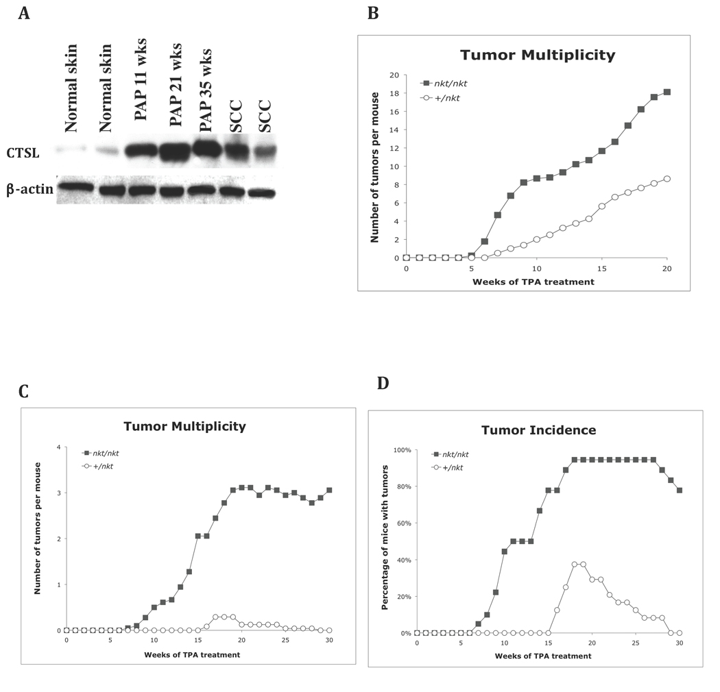
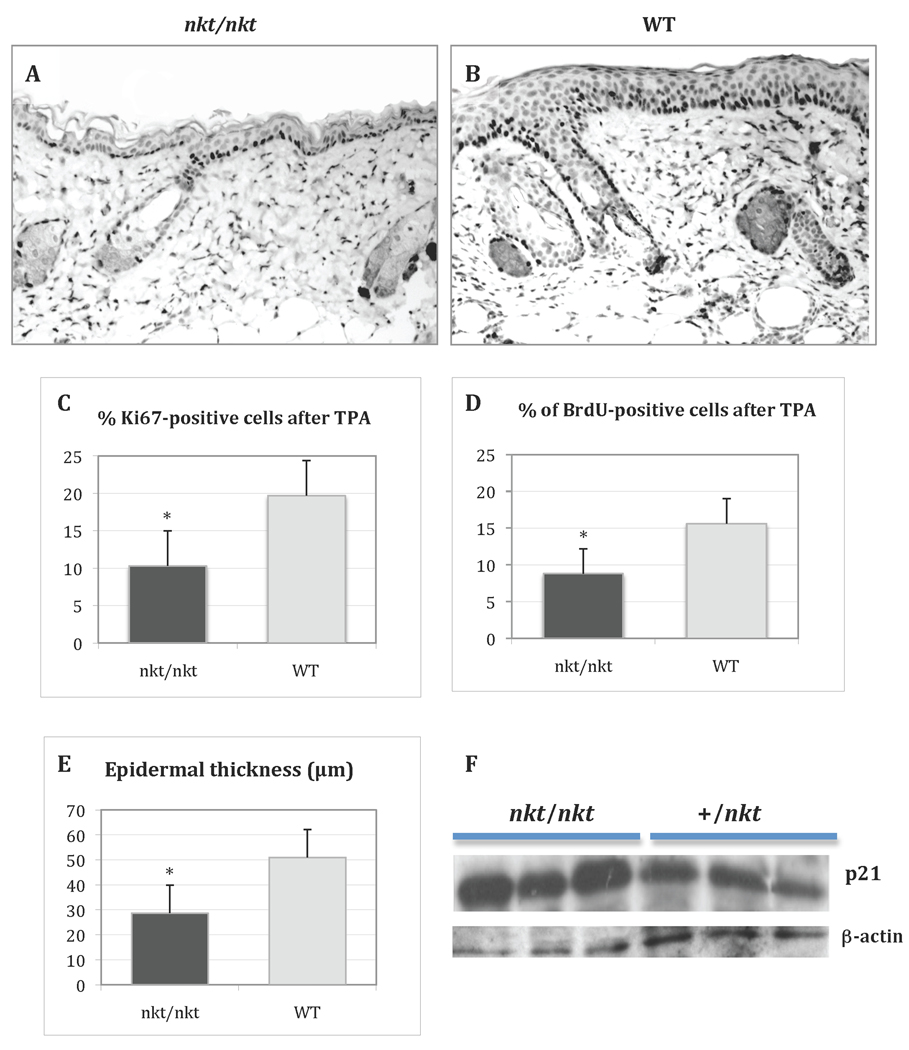
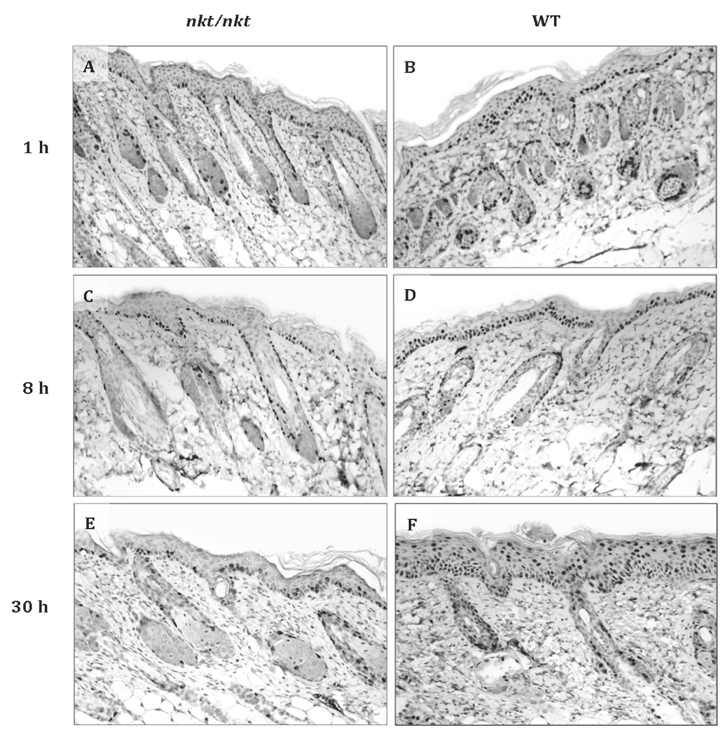
Lysosomal cysteine protease cathepsin L (CTSL) is believed to play a role in tumor progression and is considered a marker for clinically invasive tumors. Studies from our laboratory using the classical mouse skin carcinogenesis model, with 7,12-dimethyl-benz[a]anthracene (DMBA) for initiation and 12-O-tetradecanoylphorbol-13-acetate (TPA) for promotion, showed that expression of CTSL is increased in papillomas and squamous cell carcinomas (SCC). We also carried out carcinogenesis studies using Ctsl-deficient nackt (nkt) mutant mice on three different inbred backgrounds. Unexpectedly, the multiplicity of papillomas was significantly higher in Ctsl-deficient than in wild-type mice on two unrelated backgrounds. Topical applications of TPA or DMBA alone to the skin of nkt/nkt mice did not induce papillomas, and there was no increase in spontaneous tumors in nkt/nkt mice on any of the three inbred backgrounds. Reduced epidermal cell proliferation in Ctsl-deficient nkt/nkt mice after TPA treatment suggested that they are not more sensitive than wild-type mice to TPA promotion. We also showed that deficiency of CTSL delays terminal differentiation of keratinocytes, and we propose that decreased elimination of initiated cells is at least partially responsible for the increased papilloma formation in the nackt model.

Keywords: cysteine cathepsins; mouse models; proteases; skin cancer; two-stage carcinogenesis.


Figures

Figure 1
Figure 1. Two-stage carcinogenesis studies in Ctsl-deficient mice
(A) Western blot of normal skin, early (11 wks), mid (21 wks), and late (35 wks) papillomas and squamous cell carcinomas from SENCARB/Pt mice showing how CTSL protein expression increases during papilloma development (antibody: M-19, sc-6500). (B) Tumor multiplicity of skin tumors after two-stage carcinogenesis in SENCARB/Pt-nkt/nkt mice (n = 9) was statistically highly significant from that of wild-type mice (n = 7) at 20 wk (P< 0.001, Wilcoxon Rank Sum test). (C) Tumor multiplicity in DBA/2-nkt/nkt mice (n = 18) was statistically highly significant from that of wild-type mice (n = 20) at 20 wk (P< 0.001). (D) Tumor incidence in DBA/2-nkt/nkt mice reached a plateau of 94% at 18 wk compared to 38% for wild-type mice (all the papillomas from this group regressed by wk 30). For two-stage carcinogenesis, 6–8 wk old mice were initiated with DMBA and after two weeks received repeated applications of TPA as described in Materials & Methods. The number of papillomas was determined weekly. The tumor incidence is defined as the percentage of mice with skin tumors and the tumor multiplicity is the average number of skin tumors per mouse. Mutant nkt/nkt mice (ν); wild-type mice (μ).
Figure 2
Figure 2. Typical papilloma and atypical SCC with basaloid proliferation and follicular differentiation in nkt/nkt mice
Representative immunohistochemistry images of a papilloma (at 30 weeks of promotion) from Ctsl-deficient DBA/2-nkt/nkt mice induced by the two-stage carcinogenesis protocol showing BrdU-labeled nuclei (A) and the absence of CTSL expression (B). Insets show wild-type papillomas from DBA/2 mice induced by the two-stage carcinogenesis (from archived samples). Representative hematoxylin and eosin staining images of an atypical SCC from Ctsl-deficient DBA/2-nkt/nkt mice showing epithelial proliferation composed of basaloid cells and the presence of keratin cysts (arrows) (C and D). Representative immunohistochemistry staining images showing the expression of basal cell marker K14 (E) and the absence of expression for the differentiation marker K10 (F) by the basaloid cells. Magnifications at ×40 (A, B, C, E and F) and ×100 (D).
Figure 3
Figure 3. Two-stage carcinogenesis study in CD4 T-cell deficient mice
Tumor multiplicity of FVB/N-Cd4 −/− mice (n = 10) was statistically highly significant from that of FVB/N wild-type mice (n = 8) at 20 wk (P< 0.001, Wilcoxon Rank Sum test). Mice 6–8 wk old were initiated with DMBA and after two weeks received repeated applications of TPA as described in Materials & Methods. The number of papillomas was determined weekly. Tumor multiplicity is the average number of skin tumors per mouse. FVB/N wild-type mice (ν); Cd4 −/− mice (μ).
Figure 4
Figure 4. Epidermal proliferation after TPA treatment
Representative BrdU immunostaining of TPA-treated dorsal skin from a DBA/2-nkt/nkt mouse (A) and a wild-type littermate (B) (both magnifications ×100). Mice were treated with four topical applications of 3.4 nmol of TPA over a 2-wk period and sacrificed 24 h after the final treatment. Bar graphs show cell proliferation level as measured by Ki67 (C) and BrdU (D) indexes, as well as epidermal thickness (E). The determination of epidermal thickness and labeling index was performed as described in Materials and Methods. Values represent mean ± SD (*P<.05). Black bars, mutant mice; grey bars, wild-type littermates. (F) Western blot analysis of p21 in the epidermis of TPA-treated dorsal skin from DBA/2-nkt/nkt mice and littermate controls. Protein was normalized to β-actin.
Figure 5
Figure 5. Keratinocyte transit after TPA treatment
Representative BrdU immunostaining of TPA-treated dorsal skin from a DBA/2-nkt/nkt mouse and a wild-type littermate after 1 h (A and B), 8 h (C and D), and 30 h (E and F) of BrdU injection (all magnifications ×100). Mice were injected i.p. with BrdU 17 h after the last of four TPA applications (3.4 nmol) over a two week period and sacrificed 1, 8, and 30 h after BrdU injection. A total of 4 mice per genotype per time point were evaluated. At 8 h and 30 h after BrdU injection, progression of BrdU-labeled keratinocytes towards the surface is clearly slower in the mutant epidermis. Percentages of BrdU-labeled nuclei in the basal and suprabasal layer for the three time points are shown in Table 1.

References

    1. Mohamed MM, Sloane BF. Cysteine cathepsins: multifunctional enzymes in cancer. Nat Rev Cancer. 2006;6(10):764–775. - PubMed
    1. Gocheva V, Zeng W, Ke D, et al. Distinct roles for cysteine cathepsin genes in multistage tumorigenesis. Genes Dev. 2006;20(5):543–556. - PMC - PubMed
    1. Roth W, Deussing J, Botchkarev VA, et al. Cathepsin L deficiency as molecular defect of furless: hyperproliferation of keratinocytes and pertubation of hair follicle cycling. Faseb J. 2000;14(13):2075–2086. - PubMed
    1. Benavides F, Starost MF, Flores M, Gimenez-Conti IB, Guenet JL, Conti CJ. Impaired hair follicle morphogenesis and cycling with abnormal epidermal differentiation in nackt mice, a cathepsin L-deficient mutation. Am J Pathol. 2002;161(2):693–703. - PMC - PubMed
    1. Tobin DJ, Foitzik K, Reinheckel T, et al. The lysosomal protease cathepsin L is an important regulator of keratinocyte and melanocyte differentiation during hair follicle morphogenesis and cycling. Am J Pathol. 2002;160(5):1807–1821. - PMC - PubMed

Publication types

MeSH terms