Review
. 2011 Jun;91(6):511-25.
doi: 10.1002/bdra.20809. Epub 2011 Apr 28.

What chick and mouse models have taught us about the role of the endocardium in congenital heart disease

Affiliations
Review

What chick and mouse models have taught us about the role of the endocardium in congenital heart disease

Daniel M DeLaughter et al. Birth Defects Res A Clin Mol Teratol. 2011 Jun.

Abstract

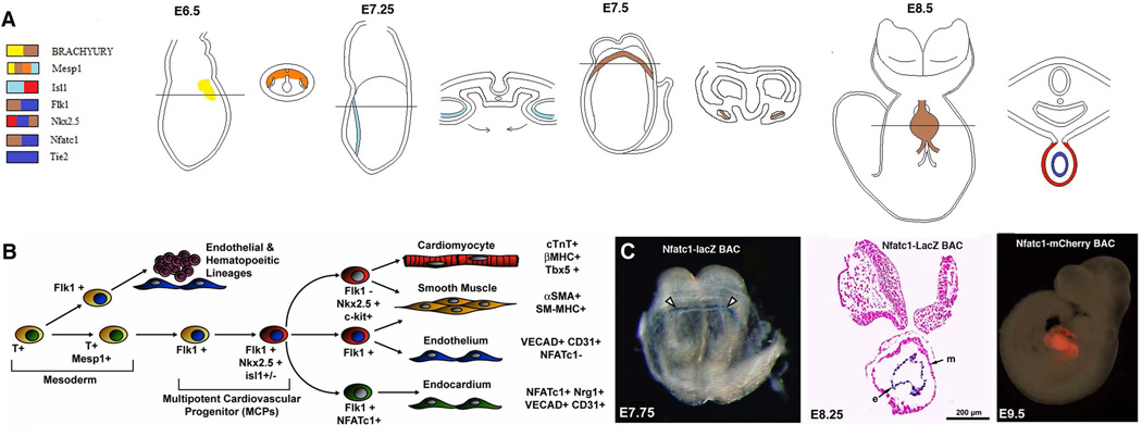
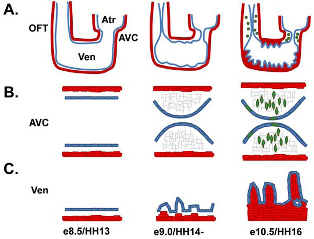
Specific cell and tissue interactions drive the formation and function of the vertebrate cardiovascular system. Although much attention has been focused on the muscular components of the developing heart, the endocardium plays a key role in the formation of a functioning heart. Endocardial cells exhibit heterogeneity that allows them to participate in events such as the formation of the valves, septation of the outflow tract, and trabeculation. Here we review, the contributions of the endocardium to cardiovascular development and outline useful approaches developed in the chick and mouse that have revealed endocardial cell heterogeneity, the signaling molecules that direct endocardial cell behavior, and how these insights have contributed to our understanding of cardiovascular development and disease.


Figures

Figure 1
Figure 1. Gene Demarcating Pro- and Definitive Endocardial Cells
(A.) The genes listed in the legend are sequentially expressed within cardiogenic precursors that will ultimately contribute to the endocardium in vivo. At E6.5, cells of the epiblast begin to ingress through the primitive streak (PS) and those of the future mesoderm will begin to express Brachyury T. This includes the population of endocardial progenitors. These cells will also co-express other mesoderm specific genes such as Mesp1. By E7.25, cells of the same population will reside in the splanchnic mesoderm and can be identified by the expression of Isl1. The cardiac crescent that is visible by E7.5 contains cells of the splanchnic mesoderm that now express Flk1, Nkx2.5, and Nfatc1. At E8.25, the inner endocardial layer of the heart tube can be detected by the expression of Nfatc1 and Tie2. Above are images of whole mount embryos at the designated stages. Aside each whole mount image is a transverse section of the embryo. (B.) The pathway illustrated provides an in vitro delineation of genes expressed specifically during endocardial development. (C.) Representative images of Nfatc1 -LacZ BAC and Nfatc1-mCherry BAC expression in vivo within cardiogenic regions of a developing embryo at specific stages. The coronal section at E8.25 marks the endocardium (e) and the myocardium (m) of the heart tube.
Figure 2
Figure 2. Endocardial functional heterogeneity during tubular heart morphogenesis
(A.) At e8.5/HH13, the u-shaped tubular heart is comprised of an outer layer of Myocardium (Red) and an inner layer of Endocardium (blue) with a layer of cardiac jelly in between. Possessing a common outflow tract (OFT), ventricle (Ven), atria (Atr) and atrioventricular canal (AVC), the region between the atria and ventricle. (B.) By e9.0/HH14- swellings of ECM at the AVC and OFT form the endocardial cushions and endocardial cell EMT is just about to begin in the AVC, while the OFT begins EMT a day later. By e10.5/HH16 endocardial cells overlaying the cushions undergo EMT and invade cardiac jelly. (C.) The ventricular endocardium does not undergo EMT, rather it begins the complicated process of trabeculation around this time. The atrial endocardium undergoes neither EMT or trabeculation at this time.
Figure 3
Figure 3. Viral gene transfer into the endocardium of the tubular heart
HH10-12 chicks are dissected into whatman rings and injected with virus expressing either GFP or a gene of interest. These embryos are incubated for 24 hours on egg agar. The atrioventricular canal (AVC) or ventricle (Ven) are excised, cut lengthwise, and explanted endocardial side down onto a collagen I gel. After 48 hours incubation, the explants are fixed and the total number of GFP positive transformed cells (in green) in the gel are counted. Loss of function can be scored as a reduction in the number of transformed cells in gels incubated with AVC explants. Gain of function scored is scored as the increase in the total number of GFP positive cells in gels incubated with Ven explants. Abbreviations: Endo-Endocardium, Myo-Myocardium, Gel-Rat tail collagen I gel.

References

    1. Aikawa E, Whittaker P, Farber M, Mendelson K, Padera RF, Aikawa M, Schoen FJ. Human semilunar cardiac valve remodeling by activated cells from fetus to adult: implications for postnatal adaptation, pathology, and tissue engineering. Circulation. 2006;113(10):1344–1352. - PubMed
    1. Akiyama H, Chaboissier MC, Behringer RR, Rowitch DH, Schedl A, Epstein JA, de Crombrugghe B. Essential role of Sox9 in the pathway that controls formation of cardiac valves and septa. Proc Natl Acad Sci U S A. 2004;101(17):6502–6507. - PMC - PubMed
    1. Araki T, Mohi MG, Ismat FA, Bronson RT, Williams IR, Kutok JL, Yang W, Pao LI, Gilliland DG, Epstein JA, Neel BG. Mouse model of Noonan syndrome reveals cell type- and gene dosage-dependent effects of Ptpn11 mutation. Nat Med. 2004;10(8):849–857. - PubMed
    1. Baldwin HS. Early embryonic vascular development. Cardiovasc Res. 1996;31:E34–E45. Spec No. - PubMed
    1. Baldwin HS, Jensen KL, Solursh M. Myogenic cytodifferentiation of the precardiac mesoderm in the rat. Differentiation. 1991;47(3):163–172. - PubMed

Publication types