Case Reports
. 2011 Jun;155A(6):1298-313.
doi: 10.1002/ajmg.a.33970. Epub 2011 Apr 28.

Identification of p.A684V missense mutation in the WFS1 gene as a frequent cause of autosomal dominant optic atrophy and hearing impairment

Affiliations
Case Reports

Identification of p.A684V missense mutation in the WFS1 gene as a frequent cause of autosomal dominant optic atrophy and hearing impairment

Nanna D Rendtorff et al. Am J Med Genet A. 2011 Jun.

Abstract

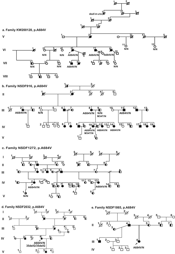
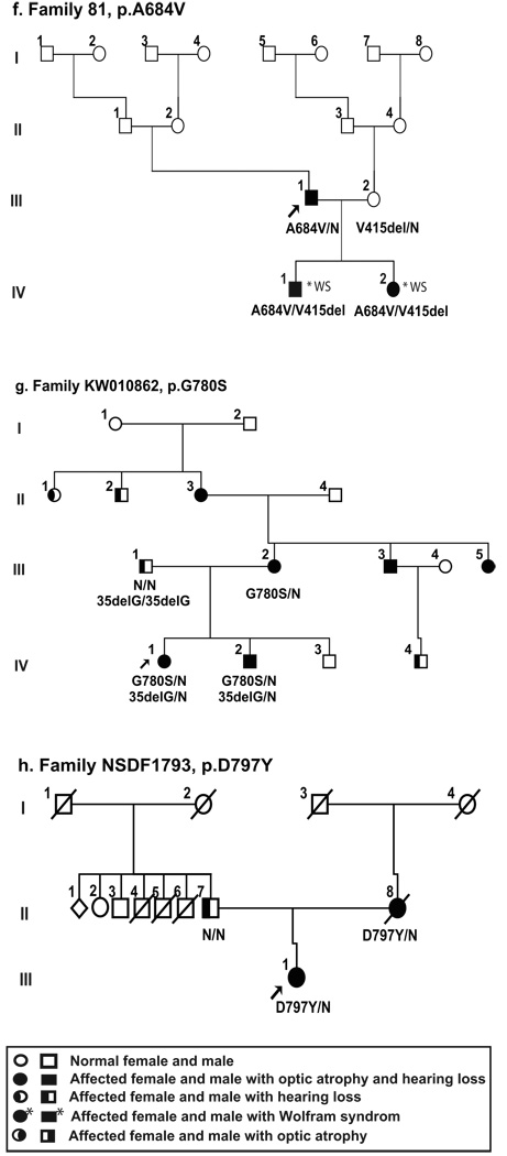
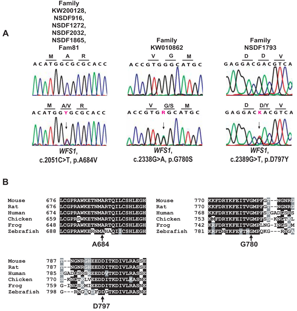
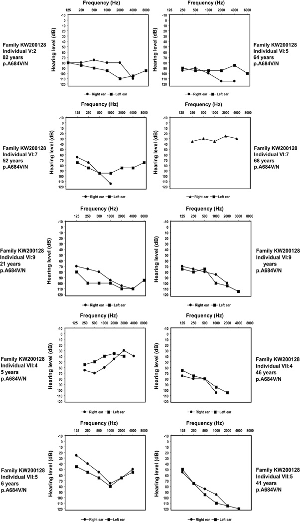
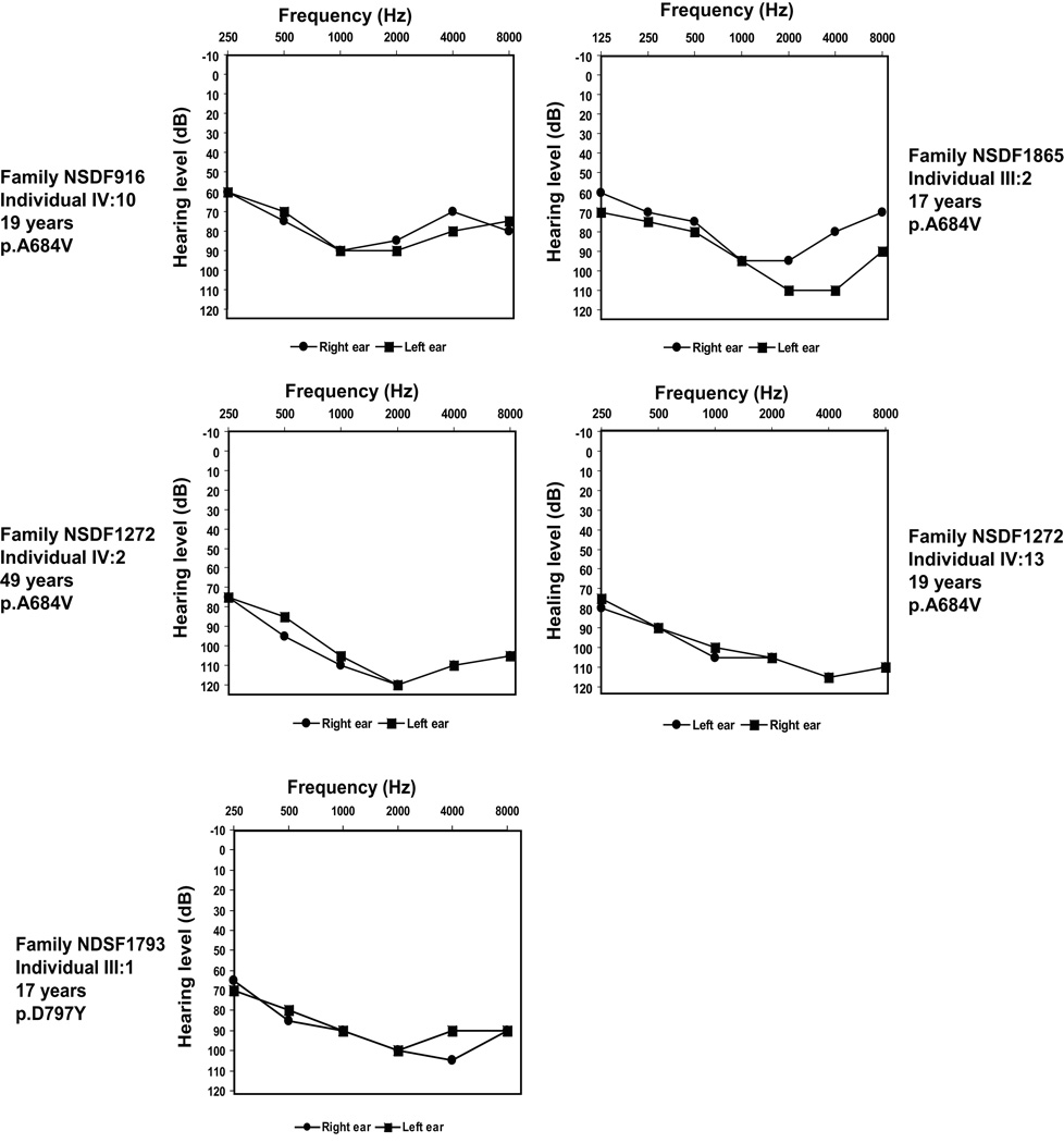
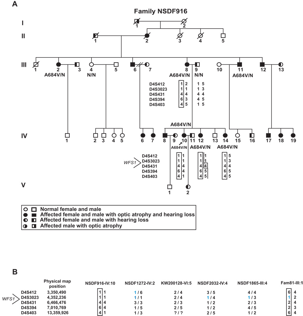
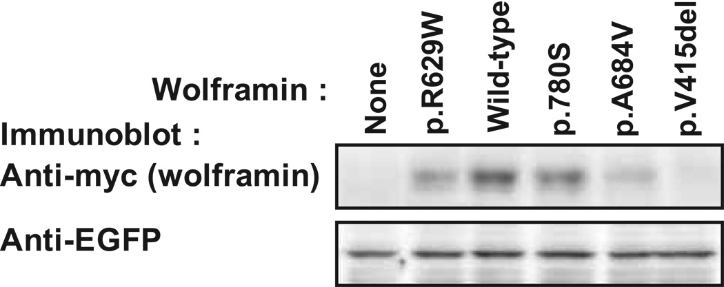
Optic atrophy (OA) and sensorineural hearing loss (SNHL) are key abnormalities in several syndromes, including the recessively inherited Wolfram syndrome, caused by mutations in WFS1. In contrast, the association of autosomal dominant OA and SNHL without other phenotypic abnormalities is rare, and almost exclusively attributed to mutations in the Optic Atrophy-1 gene (OPA1), most commonly the p.R445H mutation. We present eight probands and their families from the US, Sweden, and UK with OA and SNHL, whom we analyzed for mutations in OPA1 and WFS1. Among these families, we found three heterozygous missense mutations in WFS1 segregating with OA and SNHL: p.A684V (six families), and two novel mutations, p.G780S and p.D797Y, all involving evolutionarily conserved amino acids and absent from 298 control chromosomes. Importantly, none of these families harbored the OPA1 p.R445H mutation. No mitochondrial DNA deletions were detected in muscle from one p.A684V patient analyzed. Finally, wolframin p.A684V mutant ectopically expressed in HEK cells showed reduced protein levels compared to wild-type wolframin, strongly indicating that the mutation is disease-causing. Our data support OA and SNHL as a phenotype caused by dominant mutations in WFS1 in these additional eight families. Importantly, our data provide the first evidence that a single, recurrent mutation in WFS1, p.A684V, may be a common cause of ADOA and SNHL, similar to the role played by the p.R445H mutation in OPA1. Our findings suggest that patients who are heterozygous for WFS1 missense mutations should be carefully clinically examined for OA and other manifestations of Wolfram syndrome.


Figures

Figure 1
Figure 1
Pedigrees of the eight families investigated in this study. All the families show a segregation pattern compatible with the conclusion that the indicated WFS1 mutation causes autosomal dominant optic atrophy and hearing loss, except for family 81, where the father of two children with Wolfram syndrome is a sporadic case. Probands are indicated by arrows. Black symbols indicate optic atrophy with hearing loss patients. Black shading on the left part of symbol indicates individuals with isolated hearing loss and on the right part individuals with isolated optic atrophy. The WFS1 molecular result is indicated below individuals from whom DNA samples were available. N = normal allele. The presence of GJB2 mutation are indicated (c.35delG or p.M34T).
Figure 1
Figure 1
Pedigrees of the eight families investigated in this study. All the families show a segregation pattern compatible with the conclusion that the indicated WFS1 mutation causes autosomal dominant optic atrophy and hearing loss, except for family 81, where the father of two children with Wolfram syndrome is a sporadic case. Probands are indicated by arrows. Black symbols indicate optic atrophy with hearing loss patients. Black shading on the left part of symbol indicates individuals with isolated hearing loss and on the right part individuals with isolated optic atrophy. The WFS1 molecular result is indicated below individuals from whom DNA samples were available. N = normal allele. The presence of GJB2 mutation are indicated (c.35delG or p.M34T).
Figure 2
Figure 2
(A) Representative sequence chromatograms for each of the WFS1 missense mutations compared to a normal control. The arrows indicate the nucleotide changes of the heterozygous missense mutations. Nomenclature of mutations refers to the WFS1 RefSeq NM_006005.2, with nucleotide number +1 being A of the start codon ATG. Each mutation is heterozygous in affected individuals. (B) Alignment in different species shows strong evolutionary conservation of the relevant amino acid mutated in the patients.
Figure 3
Figure 3
Pure-tone audiograms of left and right ear of representative, affected family members heterozygous for WFS1 mutation from six of the families studies (KW2001128 (audiograms from 6 individuals), NSDF916, NSDF1865, NSDF1272, and NSDF1793). For some individuals two audiograms are shown to illustrate the hearing loss over time. Individual VI:7 (family KW200128) had cochlear implant at age 57 years, resulting in an improved hearing between age 52 and age 68.
Figure 3
Figure 3
Pure-tone audiograms of left and right ear of representative, affected family members heterozygous for WFS1 mutation from six of the families studies (KW2001128 (audiograms from 6 individuals), NSDF916, NSDF1865, NSDF1272, and NSDF1793). For some individuals two audiograms are shown to illustrate the hearing loss over time. Individual VI:7 (family KW200128) had cochlear implant at age 57 years, resulting in an improved hearing between age 52 and age 68.
Figure 4
Figure 4
Haplotype analysis. (A) Haplotype analysis in Family NSDF916, harboring the p.A684V mutation, using five polymorphic markers flanking WFS1. (B) Marker analysis with the same five markers in probands from the other five p.A684V families. A bar indicates the haplotype carrying the mutation in those families where this could be deduced.
Figure 5
Figure 5
Functional protein expression analysis of WFS1 mutations. HEK293 cells were co-transfected with plasmid expressing myc-tagged wild-type or mutant wolframin together with plasmid expressing EGFP. The cells were lysed 16 hours post-transfection and the cell extracts were subjected to SDS-PAGE and immunoblotting for the myc tag and EGFP. The experiment was repeated 3 times with independently generated mutants with similar results.

References

    1. Amati-Bonneau P, Guichet A, Olichon A, Chevrollier A, Viala F, Miot S, Ayuso C, Odent S, Arrouet C, Verny C, Calmels MN, Simard G, Belenguer P, Wang J, Puel JL, Hamel C, Malthiery Y, Bonneau D, Lenaers G, Reynier P. OPA1 R445H mutation in optic atrophy associated with sensorineural deafness. Ann Neurol. 2005;58:958–963. - PubMed
    1. Amati-Bonneau P, Milea D, Bonneau D, Chevrollier A, Ferre M, Guillet V, Gueguen N, Loiseau D, de Crescenzo MA, Verny C, Procaccio V, Lenaers G, Reynier P. OPA1-associated disorders: phenotypes and pathophysiology. Int J Biochem Cell Biol. 2009;41:1855–1865. - PubMed
    1. Amati-Bonneau P, Odent S, Derrien C, Pasquier L, Malthiery Y, Reynier P, Bonneau D. The association of autosomal dominant optic atrophy and moderate deafness may be due to the R445H mutation in the OPA1 gene. Am J Ophthalmol. 2003;136:1170–1171. - PubMed
    1. Amati-Bonneau P, Valentino ML, Reynier P, Gallardo ME, Bornstein B, Boissiere A, Campos Y, Rivera H, de la Aleja JG, Carroccia R, Iommarini L, Labauge P, Figarella-Branger D, Marcorelles P, Furby A, Beauvais K, Letournel F, Liguori R, La MC, Montagna P, Liguori M, Zanna C, Rugolo M, Cossarizza A, Wissinger B, Verny C, Schwarzenbacher R, Martin MA, Arenas J, Ayuso C, Garesse R, Lenaers G, Bonneau D, Carelli V. OPA1 mutations induce mitochondrial DNA instability and optic atrophy 'plus' phenotypes. Brain. 2008;131:338–351. - PubMed
    1. Arnos KS, Welch KO, Tekin M, Norris VW, Blanton SH, Pandya A, Nance WE. A comparative analysis of the genetic epidemiology of deafness in the United States in two sets of pedigrees collected more than a century apart. Am J Hum Genet. 2008;83:200–207. - PMC - PubMed

Publication types

MeSH terms