. 2011 May 31;50(21):4479-90.
doi: 10.1021/bi2003907. Epub 2011 May 3.

Effect of glycans and the glycophosphatidylinositol anchor on strain dependent conformations of scrapie prion protein: improved purifications and infrared spectra

Affiliations

Effect of glycans and the glycophosphatidylinositol anchor on strain dependent conformations of scrapie prion protein: improved purifications and infrared spectra

Gerald S Baron et al. Biochemistry. .

Abstract

Mammalian prion diseases involve conversion of normal prion protein, PrP(C), to a pathological aggregated state (PrP(res)). The three-dimensional structure of PrP(res) is not known, but infrared (IR) spectroscopy has indicated high, strain-dependent β-sheet content. PrP(res) molecules usually contain a glycophosphatidylinositol (GPI) anchor and large Asn-linked glycans, which can also vary with strain. Using IR spectroscopy, we tested the conformational effects of these post-translational modifications by comparing wild-type PrP(res) with GPI- and glycan-deficient PrP(res) produced in GPI-anchorless PrP transgenic mice. These analyses required the development of substantially improved purification protocols. Spectra of both types of PrP(res) revealed conformational differences between the 22L, ME7, and Chandler (RML) murine scrapie strains, most notably in bands attributed to β-sheets. These PrP(res) spectra were also distinct from those of the hamster 263K scrapie strain. Spectra of wild-type and anchorless 22L PrP(res) were nearly indistinguishable. With ME7 PrP(res), modest differences between the wild-type and anchorless spectra were detected, notably an ∼2 cm(-1) shift in an apparent β-sheet band. Collectively, the data provide evidence that the glycans and anchor do not grossly affect the strain-specific secondary structures of PrP(res), at least relative to the differences observed between strains, but can subtly affect turns and certain β-sheet components. Recently reported H-D exchange analyses of anchorless PrP(res) preparations strongly suggested the presence of strain-dependent, solvent-inaccessible β-core structures throughout most of the C-terminal half of PrP(res) molecules, with no remaining α-helix. Our IR data provide evidence that similar core structures also comprise wild-type PrP(res).


Figures

Figure 1
Figure 1
Ultrastructure of anchorless PrPres fibrils. Samples were stained with methylamine tungstate and examined by TEM. Images of mock anchorless PrPres, buffer, and purified mouse ferritin are shown as controls. Arrows, ferritin particles. Bar, 100 nm.
Figure 2
Figure 2
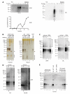
Biochemical characterization of anchorless PrPres preparations. (A) Silver-stained SDS-PAGE gels. Varying amounts of hamster rPrPsen 90-231 (ng) were loaded as indicated (lanes 1-3). Lanes 8-9 and 10-11 each represent samples from independent preparations. Bracket indicates bands corresponding to anchorless PrPres monomers. (B) Immunoblot analysis with 6D11 or R20 anti-PrP antibodies. Varying amounts of hamster rPrPsen 90-231 (ng) were loaded as indicated (lanes 1-6). Brackets (lower panels) indicate strain-specific low molecular mass species. (C) Immunoblot with anti-heavy chain ferritin antibody. Blots were performed using antibody without (Untreated) or with (Ferritin pre-adsorbed) pre-adsorption with purified mouse ferritin to determine ferritin-specific antibody binding. Varying amounts of purified mouse liver ferritin (ng) were loaded as indicated (lanes 1-3 and 7-9). GPIneg PrPres, two independent preparations of anchorless 22L PrPres containing either 1.2 (lanes 4 and 10) or 2 μg (lanes 5 and 11) of PrPres. wt std PrPres, 300 ng of wt 22L PrPres prepared by a standard method (41). For (A)-(C), molecular mass standards are indicated on the left (kDa) and white lines indicate removal of irrelevant lanes.
Figure 3
Figure 3
Biochemical characterization of wt PrPres preparations. (A) PrPres enriched in brain DRM fractions. Immunoblot analysis (with 6D11 antibody) of PK-treated fractions from brain DRM preparations from hamsters (263K) or mice (22L) infected with rodent scrapie. Arrows indicate DRM band fractions (fractions 5-6). Fractions 2-3 correspond to a second major lipid band of lower buoyant density observed in the gradient. Graph shows distribution of total protein across fractions from 263K brain DRM gradient. (B) Silver-stain SDS-PAGE gel analysis of PrPres preparations from hamsters and mice using the standard method (std) versus the DRM method (DRM). Similar amounts of std and DRM PrPres were loaded based on PrP content. Control mock preparations from hamsters or mice are shown for comparison. Std and DRM mock PrPres samples (lanes 3-4 and 7-8) were normalized to each other based on brain equivalents. Arrow, ferritin band present in mock and scrapie PrPres preparations using the standard method. Brackets, low molecular mass species that are significantly reduced in DRM PrPres preparations. (C) Immunoblot analysis (6D11 antibody) of PrPres preparations. Varying amounts of std PrPres (ng) were loaded as indicated (lanes 1-3 and 6-9). Arrows indicate the three major glycoforms of PrP. Brackets, SDS-resistant PrPres oligomers. (D) Immunoblots of 263K PrPres with anti-PrP (R20) and anti-heavy chain ferritin antibodies. Varying amounts of std 263K PrPres (ng) were loaded as indicated. Ferritin was not detected in the DRM PrPres prep (DRM) but readily detected in a std PrPres prep with comparable loading for PrP (arrows). (E) Immunoblot of 22L PrPres with anti-heavy chain ferritin antibody. Dilution series of purified mouse ferritin without (lanes 1-4) or with PK treatment (7-10) were loaded as indicated. Lanes 5 and 6 contain 150 ng of either std or DRM 22L PrPres, respectively. Arrow, ferritin monomer. SDS-resistant ferritin oligomers are also visible. For (A)-(E), molecular mass standards are indicated on the left (kDa) and white lines indicate removal of irrelevant lanes.
Figure 4
Figure 4
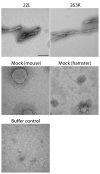
Ultrastructure of wt DRM PrPres fibrils. Samples were stained with ammonium molybdate and examined by TEM. Grids of mock DRM PrPres from mice or hamsters were exposed to 10-fold more brain equivalents than 263K or 22L PrPres grids to compensate for the extremely low levels of material in mock PrPres samples. Bar, 100 nm.
Figure 5
Figure 5
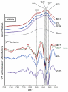
IR spectra of anchorless PrPres (murine ME7, Chandler, and 22L strains) and recombinant PrPsen 90-231. Individual primary (top) and second derivative (bottom) IR spectra represent independent preparations. For comparison, a spectrum of comparable brain equivalents of a mock anchorless PrPres preparation from normal brain tissue is shown. The spectra of the PrPres preparations have been normalized to one another so that the absorbance terms are arbitrary. The Chandler spectra are the same as those presented elsewhere in a different context as Supplemental Data (42).
Figure 6
Figure 6
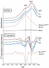
IR spectra of wild type PrPres (mouse ME7 and 22L, hamster 263K strains). Individual primary (top) and second derivative (bottom) spectra represent independent preparations. Comparable brain equivalent of a mock wild type PrPres preparation from normal brain tissue is shown as well as a second derivative spectrum calculated from an ME7-mock difference spectrum (green). Compared to the PrPres spectra, the mock spectrum also displayed a much reduced relative absorbance in the protein amide II region (~1545 cm−1, not shown) suggesting that the absorbance at ~1650 cm−1 is due predominantly to something other than protein. The spectra of the PrPres preparations have been normalized to one another so that the absorbance terms are arbitrary.
Figure 7
Figure 7
Comparisons of anchorless and wild type PrPres second derivative IR spectra.

References

    1. Pan K-M, Baldwin M, Nguyen J, Gasset M, Serban A, Groth D, Mehlhorn I, Huang Z, Fletterick RJ, Cohen FE, Prusiner SB. Conversion of alpha-helices into beta-sheets features in the formation of the scrapie prion protein. Proc. Natl. Acad. Sci. USA. 1993;90:10962–10966. - PMC - PubMed
    1. Riek R, Hornemann S, Wider G, Glockshuber R, Wuthrich K. NMR characterization of the full-length recombinant murine prion protein, mPrP(23-231) FEBS Lett. 1997;413:282–288. - PubMed
    1. Riek R, Hornemann S, Wider G, Billeter M, Glockshuber R, Wuthrich K. NMR structure of the mouse prion protein domain PrP(121-231) Nature. 1996;382:180–182. - PubMed
    1. Caughey BW, Dong A, Bhat KS, Ernst D, Hayes SF, Caughey WS. Secondary structure analysis of the scrapie-associated protein PrP 27-30 in water by infrared spectroscopy. Biochemistry. 1991;30:7672–7680. - PubMed
    1. Gasset M, Baldwin MA, Fletterick RJ, Prusiner SB. Perturbation of the secondary structure of the scrapie prion protein under conditions that alter infectivity. Proc. Natl. Acad. Sci. USA. 1993;90:1–5. - PMC - PubMed

Publication types