. 2011 Jun 27;51(6):1393-404.
doi: 10.1021/ci200077t. Epub 2011 May 13.

Interactions of halichondrin B and eribulin with tubulin

Affiliations

Interactions of halichondrin B and eribulin with tubulin

Ruoli Bai et al. J Chem Inf Model. .

Abstract

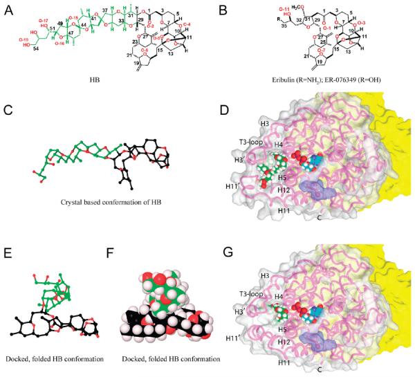
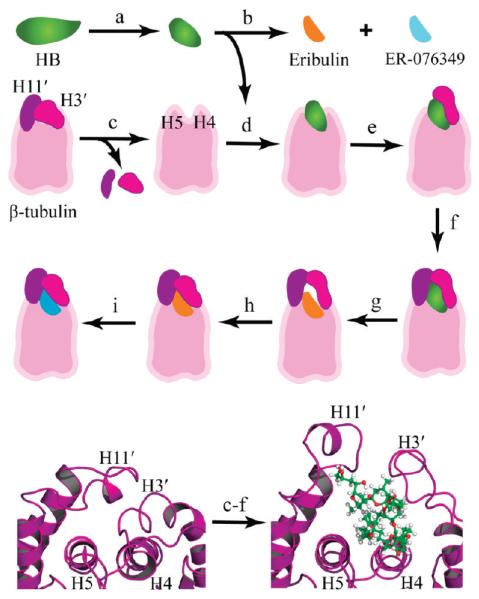
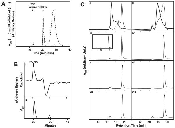
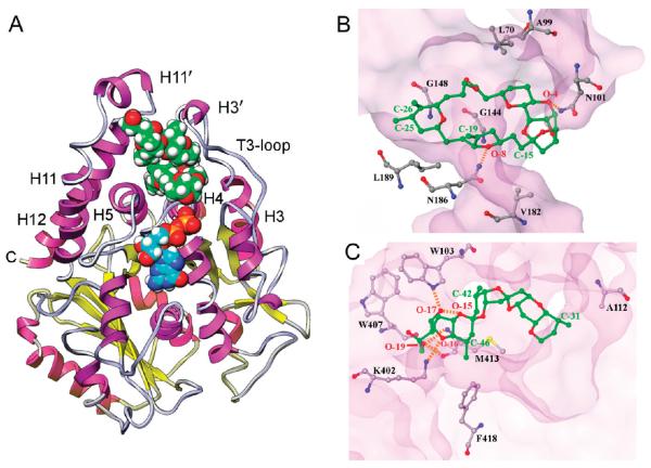
Compounds that modulate microtubule dynamics include highly effective anticancer drugs, leading to continuing efforts to identify new agents and improve the activity of established ones. Here, we demonstrate that [(3)H]-labeled halichondrin B (HB), a complex, sponge-derived natural product, is bound to and dissociated from tubulin rapidly at one binding site per αβ-heterodimer, with an apparent K(d) of 0.31 μM. We found no HB-induced aggregation of tubulin by high-performance liquid chromatography, even following column equilibration with HB. Binding of [(3)H]HB was competitively inhibited by a newly approved clinical agent, the truncated HB analogue eribulin (apparent K(i), 0.80 μM) and noncompetitively by dolastatin 10 and vincristine (apparent K(i)'s, 0.35 and 5.4 μM, respectively). Our earlier studies demonstrated that HB inhibits nucleotide exchange on β-tubulin, and this, together with the results presented here, indicated the HB site is located on β-tubulin. Using molecular dynamics simulations, we determined complementary conformations of HB and β-tubulin that delineated in atomic detail binding interactions of HB with only β-tubulin, with no involvement of the α-subunit in the binding interaction. Moreover, the HB model served as a template for an eribulin binding model that furthered our understanding of the properties of eribulin as a drug. Overall, these results established a mechanistic basis for the antimitotic activity of the halichondrin class of compounds.


Figures

Figure 1
Figure 1
Molecular structures of HB, eribulin, and ER-076349 and the binding poses of HB and eribulin on β-tubulin. (A) Structure of HB (color code in E/F). (B) Structures of eribulin and ER-076349. (C) Ball-and-stick structure of HB in a conformation based on the crystal structure of NBE (color code in E/F). (D) HB bound to β-tubulin. The surface of β-tubulin is gray (α-tubulin is yellow), except for the portion colored purple that interacts with vinblastine in the 1Z2B structure. The β-tubulin polypeptide chain is shown as a pink strand, with key features indicated. The E-site GDP and HB are shown with cyan and green carbons, respectively. Other atoms: hydrogen, white; oxygen, red; nitrogen, blue; and phosphorus, orange. (E and F) Folded conformation of HB, obtained from docking studies presented here, rendered in ball-and-stick and Corey–Pauling–Koltun (CPK), respectively. Carbon atoms of the side chain are green to illustrate the major conformational change HB undergoes from the derived crystal structure to the bound conformation. Macrocycle carbon atoms are black, and oxygen atoms are red. The tight packing of the HB side chain on top of the macrocycle demonstrates the hydrophobic collapse in the folded conformations, so that solvent exposure of HB is minimized. Hydrogen atoms (light lavender) are shown in the CPK diagram. (G) Eribulin bound to β-tubulin. Details as in panel D.
Figure 2
Figure 2
Modeling the HB binding site. Top three rows: stepwise approach used to model the binding modes. (a) The crystal-based structure of HB was subjected to molecular dynamics, and folded conformations were selected for docking studies. (b) Eribulin and ER-076349 structures were generated from the HB folded conformations. (c) H3′ and H11′ loops were removed from a β-tubulin model to unmask the HB binding site for ligand docking. (d) HB was docked and refined. (e) The H3′ loop was reattached to β-tubulin, and the protein–ligand model was refined. (f) The H11′ loop was reattached to β-tubulin, and the atom–atom interactions were optimized to yield the HB binding model. (g) Eribulin was docked based on superimposition of the common macrocycle shared with HB. (h) Constrained simulations were used to fold the H3′ and H11′ loops onto eribulin. (i) ER-076349 was docked. Bottom row: cartoon rendering showing the key secondary structures of the HB binding site and the conformational changes associated with HB binding. The β-tubulin is rendered in purple ribbon, and HB is shown in ball-and-stick with carbon, oxygen, and hydrogen atoms colored green, red, and white, respectively. Left panel: The protein fold in the 1JFF β-tubulin structure shows the packing of the H3′ and H11′ loops onto helices H4 and H5, which occludes the HB binding site. Right panel: The protein fold in the HB binding model shows the conformational switch of the H3′ and H11′ loops upon HB binding.
Figure 3
Figure 3
HB binds to 100 kDa αβ-heterodimer. (A) Nonequilibrium binding of [3H]HB to tubulin. Dashed line, radiolabel; dotted line, A280. A 0.1 mL sample containing 5 μM each of tubulin and [3H]HB was injected into a single Shodex KW-803 column with a guard column. Running buffer: 0.1 M Mes –0.5 mM MgCl2 at 0.5 mL/min. First absorbance peak (12 min) is at the void volume (denatured tubulin aggregates). Second peak (20 min) emerges at 100 kDa. Third peak (29 min) contains unbound small molecule(s). (B) HPLC Hummel–Dreyer evaluation of the binding of [3H]HB to tubulin. (i) Radiolabel. (ii) A280. Chromatography as in panel A. Equilibration with 1.0 μM [3H]HB. Injected sample: 50 μg tubulin (0.1 mL). (C) Differences in nonequilibrium versus equilibrium HPLC gel filtration occur with vinblastine or D10 but not HB, spongistatin 1, or maytansine. Two Shodex KW-803 columns with a guard column were used in series. Flow rate: 1.0 mL/min. Injection sample: 0.1 mL, containing 10 μM tubulin. “Unequilibrated columns” means the column system had been equilibrated with 0.1 M Mes–0.5 mM MgCl2; “equilibrated columns” means the columns had been equilibrated with the buffer containing the indicated drug. Drug concentration, except with D10 (panel ii), was 20 μM in the injected sample and when the column system was equilibrated with drug (darker curve in i; panels iv, vi, and viii). The D10 concentration was 3 μM in the injected samples and equilibrated columns (panel ii, darker curve). (i) Vinblastine. Unequilibrated columns, lighter curve. Equilibrated columns, darker curve. (ii) D10. Unequilibrated columns, lighter curve. Equilibrated columns, darker curve. (iii) HB, unequilibrated columns. Inset: protein elution profile following injection of tubulin only into unequilibrated columns. (iv) HB, equilibrated columns. (v) Spongistatin 1, unequilibrated columns. (vi) Spongistatin 1, equilibrated columns. (vii) Maytansine, unequilibrated columns. (viii) Maytansine, equilibrated columns.
Figure 4
Figure 4
Hanes plots of inhibition of the binding of [3H]HB to tubulin by (A) eribulin, (B) vincristine, or (C) D10. Each 0.3 mL reaction mixture contained 2.5 μM tubulin and the indicated concentrations of [3H]HB and inhibitor. Reaction mixtures were incubated 30 min. (A) Reaction mixtures contained eribulin:○, none; Δ, 1 μM;▽, 2 μM; □, 3 μM; ◇, 4 μM. (B) Reaction mixtures contained vincristine:○, none; Δ, 3 μM;▽, 9 μM; □, 12 μM. (C) Reaction mixtures contained D10: o, none; Δ, 2 μM; ▽, 3 μM; □, 4 μM; ◇, 6 μM.
Figure 5
Figure 5
(A) HB binding site in β-tubulin. β-tubulin, rendered in ribbon, with helices in magenta, sheets in yellow, and loops in gray. E-site GDP and HB are drawn in CPK. Nitrogen, oxygen, phosphate, and hydrogen atoms are blue, red, orange, and white, respectively. The carbon atoms of HB are green; those of GDP are cyan. The HB site is at the junction of a helix-rich tertiary structure adjacent to the E-site. (B and C) Crucial HB interactions at the binding site involving the macrocycle and side chain, respectively. β-tubulin is rendered as a transparent surface, with important amino acid residues labeled and rendered in ball-and-stick, as is the (B) HB macrocycle and the (C) HB side chain. Oxygen atoms are red, nitrogen blue, and sulfur yellow. Residue carbons of β-tubulin are gray and HB carbons green. HB atom positions are labeled as in Figure 1A. Hydrogen bonds are indicated by orange dashed lines.
Figure 6
Figure 6
(A) Eribulin binding site in β-tubulin. β-tubulin structure and the CPK structures of eribulin and GDP are shown as described in the Figure 5 legend. (B and C) Crucial eribulin interactions at the binding site. Panel C also shows new intra-β-tubulin hydrogen bonds (orange dashed lines) introduced by refolding the protein over the macrocycle.
Figure 7
Figure 7
Calculated intramolecular hydrophobic-polar interaction terms for the 1000 collected conformations of eribulin and ER-076349 during their simulated binding to β-tubulin. In frame 1, each ligand starts at a position that is 20 Å away from its docked pose in the binding site on β-tubulin, and in frame 1000, each ligand assumes its docked pose in the binding pocket at a position described as 0 Å. Based on the HINT program, the hydrophobic-polar term is characterized by a negative value. Higher negative values indicate a larger magnitude of unfavorable atom-to-atom interactions.

References

    1. Bai R, Paull KD, Herald CL, Malspeis L, Pettit GR, Hamel E. Halichondrin B and homohalichondrin B, marine natural products binding in the vinca domain of tubulin: discovery of tubulin-based mechanism of action by analysis of differential cytotoxicity data. J. Biol. Chem. 1991;266:15882–15889. - PubMed
    1. Dabydeen DA, Burnett JC, Bai R, Verdier-Pinard P, Hickford SJH, Pettit GR, Blunt JW, Munro MHG, Gussio R, Hamel E. Comparison of the activities of the truncated halichondrin B analog NSC 707389 (E7389) with those of the parent compound and a proposed binding site on tubulin. Mol. Pharmacol. 2006;70:1866–1875. - PubMed
    1. Aicher TD, Buszek KR, Fang FG, Forsyth CJ, Jung SH, Kishi Y, Matelich MC, Scola PM, Spero DM, Yoon SK. Total synthesis of halichondrin B and norhalichondrin B. J. Am. Chem. Soc. 1992;114:3162–3164.
    1. Choi HW, Demeke D, Kang FA, Kishi Y, Nakajima K, Nowak P, Wan ZK, Xie CY. Synthetic studies on the marine natural product halichondrins. Pure Appl. Chem. 2003;75:1–17.
    1. Zheng W, Seletsky BM, Palme MH, Lydon PJ, Singer LA, Chase CE, Lemelin CA, Shen YC, Davis H, Tremblay L, Towle MJ, Salvato KA, Wels BF, Aalfs KK, Kishi Y, Littlefield BA, Yu MJ. Macrocyclic ketone analogues of halichondrin B. Bioorg. Med. Chem. Lett. 2004;14:5551–5554. - PubMed

Publication types