. 2011 Jul-Aug;1811(7-8):452-9.
doi: 10.1016/j.bbalip.2011.04.005. Epub 2011 Apr 22.

Interaction of enterocyte FABPs with phospholipid membranes: clues for specific physiological roles

Affiliations

Interaction of enterocyte FABPs with phospholipid membranes: clues for specific physiological roles

Lisandro J Falomir-Lockhart et al. Biochim Biophys Acta. 2011 Jul-Aug.

Abstract

Intestinal and liver fatty acid binding proteins (IFABP and LFABP, respectively) are cytosolic soluble proteins with the capacity to bind and transport hydrophobic ligands between different sub-cellular compartments. Their functions are still not clear but they are supposed to be involved in lipid trafficking and metabolism, cell growth, and regulation of several other processes, like cell differentiation. Here we investigated the interaction of these proteins with different models of phospholipid membrane vesicles in order to achieve further insight into their specificity within the enterocyte. A combination of biophysical and biochemical techniques allowed us to determine affinities of these proteins to membranes, the way phospholipid composition and vesicle size and curvature modulate such interaction, as well as the effect of protein binding on the integrity of the membrane structure. We demonstrate here that, besides their apparently opposite ligand transfer mechanisms, both LFABP and IFABP are able to interact with phospholipid membranes, but the factors that modulate such interactions are different for each protein, further implying different roles for IFABP and LFABP in the intracellular context. These results contribute to the proposed central role of intestinal FABPs in the lipid traffic within enterocytes as well as in the regulation of more complex cellular processes.


Conflict of interest statement

The authors declare no conflicts of interest.

Figures
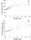
Figure 1
Figure 1. Intestinal FABPs Competition with Cytochrome c for Binding to Anionic Vesicles of Different Composition
Binding of Cytochrome c (1 μM) to PS- (Panel A) and CL-containing SUVs (Panel B) preincubated with increasing concentrations (1-12 μM) of apo-LFABP (grey circles) and apo-IFABP (black circles). Results show that both intestinal apo-FABPs interact with similar affinities and compete with cytochrome c for anionic membranes. Results are the averages ± SD of at least 3 measurements. The line represents the fitted model (see supplementary material for details). Point to point differences were estimated by Student t-Test (*p<0.05).
Figure 2
Figure 2. Binding of intestinal FABPs to sucrose loaded LUVs
The stable binding of apo-FABPs (5 μM) to LUVs (0-2 mM) of 100% EPC or 25% CL was analyzed by ultracentrifuge sedimentation. Panel A shows, as an example, the decrease of free IFABP remaining in the supernatant as the total phospholipid concentration increases. The same was observed for LFABP. The gels were quantified by densitometry and fitted individually. A representative experiment of four repetitions is shown in panel B: wild type IFABP (triangles) and IFABP-HL (square). Panel C presents the results for the parallel analysis of LFABP (circles).
Figure 3
Figure 3. Membrane destabilization by intestinal FABPs
Induced Tb/DPA complex leakage from SUVs (0.5 mM) of different composition were analyzed upon mixing with FABPs (10 μM) (apo-forms). The final leakage, expressed as % of Reference (0.05% Triton X-100), for LFABP, IFABP, IFABP-HL and αLβIFABP are shown. Statistics was based on Student t-Test (p<0.05), * indicate significant difference between EPC 100% and CL25% for the same protein; while different letters indicate differences between proteins for the same SUV type.
Figure 4
Figure 4. Hydrophobic photolabeling of intestinal FABPs
Physical interaction of native IFABP and LFABP was evidenced by radiolabeling with the photoactivable probe 125I-TID-PC employing LUVs (0.5 mM) of different compositions: 100% EPC, 25% PS and 25% CL. For each lipid composition, the upper panel shows the SDS-PAGE stained with Coomasie blue and the bottom panel its autoradiography. Results are shown here from a representative experiment. (A) shows the native proteins analyzed for the effect of the presence of oleic acid (apo- vs. holo-forms), (B) shows wild type intestinal apo-FABPs and IFABP-HL in the apo-form.
Figure 5
Figure 5. Selective proteolysis of radiolabeled wild type IFABP
After hydrophobic photolabeling with 125I-TID-PC, apo-IFABP was blotted and subjected to selective proteolisys with BNPS-Skatole. The results showed a preferential radiolabeling (right panel) of the fragment containing the α-helical region (9.6 kDa) than the one corresponding to the second half of the β-barrel (5.5 kDa) compared to the coomasie blue staining (left panel).

Similar articles

Cited by

References

    1. Bass NM. Function and Regulation of Hepatic and Intestinal Fatty-Acid Binding Proteins. Chem Phys Lipids. 1985;38:95–114. - PubMed
    1. Glatz JFC, vanderVusse GJ. Cellular fatty acid-binding proteins: Their function and physiological significance. Prog Lipid Res. 1996;35:243–282. - PubMed
    1. Storch J, Corsico B. The emerging functions and mechanisms of mammalian fatty acid-binding proteins. Annu Rev Nutr. 2008;28:73–95. - PubMed
    1. Storch J, Thumser AEA. The fatty acid transport function of fatty acid-binding proteins. Biochim Biophys Acta-Molecular and Cell Biology of Lipids. 2000;1486:28–44. - PubMed
    1. Storch J, Herr FM, Hsu KT, Kim HK, Liou HL, Smith ER. The role of membranes and intracellular binding proteins in cytoplasmic transport of hydrophobic molecules: Fatty acid-binding proteins. Comp Biochem Physiol Biochem Mol Biol. 1996;115:333–339.

Publication types

LinkOut - more resources