. 2011 Aug;91(1):335-45.
doi: 10.1016/j.yexmp.2011.04.006. Epub 2011 Apr 22.

C5b-9-activated, K(v)1.3 channels mediate oligodendrocyte cell cycle activation and dedifferentiation

Affiliations

C5b-9-activated, K(v)1.3 channels mediate oligodendrocyte cell cycle activation and dedifferentiation

Cosmin A Tegla et al. Exp Mol Pathol. 2011 Aug.

Abstract

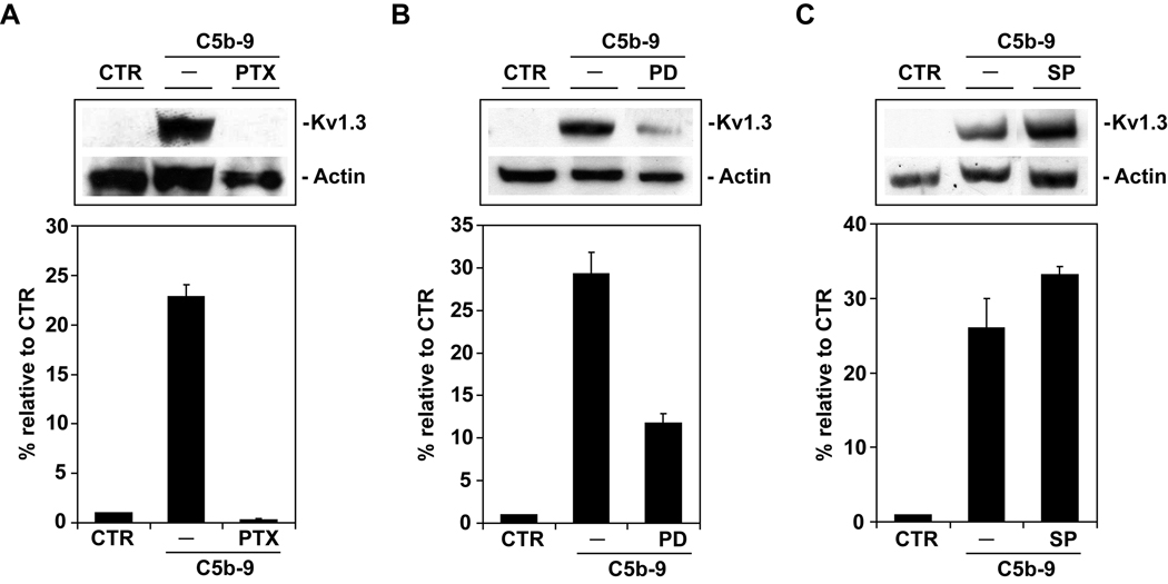
Voltage-gated potassium (K(v)) channels play an important role in the regulation of growth factor-induced cell proliferation. We have previously shown that cell cycle activation is induced in oligodendrocytes (OLGs) by complement C5b-9, but the role of K(v) channels in these cells had not been investigated. Differentiated OLGs were found to express K(v)1.4 channels, but little K(v)1.3. Exposure of OLGs to C5b-9 modulated K(v)1.3 functional channels and increased protein expression, whereas C5b6 had no effect. Pretreatment with the recombinant scorpion toxin rOsK-1, a highly selective K(v)1.3 inhibitor, blocked the expression of K(v)1.3 induced by C5b-9. rOsK-1 inhibited Akt phosphorylation and activation by C5b-9 but had no effect on ERK1 activation. These data strongly suggest a role for K(v)1.3 in controlling the Akt activation induced by C5b-9. Since Akt plays a major role in C5b-9-induced cell cycle activation, we also investigated the effect of inhibiting K(v)1.3 channels on DNA synthesis. rOsK-1 significantly inhibited the DNA synthesis induced by C5b-9 in OLG, indicating that K(v)1.3 plays an important role in the C5b-9-induced cell cycle. In addition, C5b-9-mediated myelin basic protein and proteolipid protein mRNA decay was completely abrogated by inhibition of K(v)1.3 expression. In the brains of multiple sclerosis patients, C5b-9 co-localized with NG2(+) OLG progenitor cells that expressed K(v)1.3 channels. Taken together, these data suggest that K(v)1.3 channels play an important role in controlling C5b-9-induced cell cycle activation and OLG dedifferentiation, both in vitro and in vivo.


Figures

Figure 1
Figure 1. MBP, PLP, and Kv1.3 mRNA expression during OLG differentiation
OPCs were cultured in defined medium for the indicated times, and the expression of Kv1.3, Kv 1.4, MBP, and PLP mRNA was determined. A. Expression of MBP and PLP mRNA, as determined by real-time PCR, significantly increased during OLG differentiation. B. Kv1.3 mRNA was present in the OPC cells (0 h), and the levels decreased significantly at 48h of differentiation (p=0.0007). C. The effect of differentiation on Kv protein expression was examined by western blotting and expressed as a ratio to β-actin. High levels of Kv1.3 protein were found in OPCs, and these levels decreased significantly during differentiation (p=0.029). The levels of Kv1.4 did not change significantly during differentiation. The expression of the mRNA or protein at the beginning of the experiment (0 h) was considered to be 100%. Results of three separate experiments are expressed as mean ± SEM, relative to the value at 0 h.
Figure 2
Figure 2. Effect of C5b-9 on Kv1.3 expression in OLGs
A. C5b-9 induces Kv1.3 expression. OLGs were cultured in defined medium and exposed to C5b-9 or C5D, and Kv1.3 expression was determined by western blotting. Kv1.3 protein expression was found to be low in the differentiated OLGs (CTR) and was significantly increased after exposure to C5b-9 (p=0.042). C5D serum had no significant effect on Kv1.3 expression, indicating that the increase seen was dependent on the assembly of the terminal pathway. The results are expressed as means ± SEM. Data are derived from three separate experiments. B. rOsK -1 inhibits Kv1.3 expression induced by C5b-9. OLGs were pretreated with rOsk-1 (15nM) for 1 h and then exposed to C5b-9 for 30 min. Lysates were analyzed by western blotting for Kv1.3 and β-actin expression. Exposure to rOsk-1 was able to reverse the effect of C5b-9 on Kv1.3 expression (p=0.007), indicating that channel expression is required for the C5b-9 effect. Results of three separate experiments are expressed as mean ± SEM, relative to the control (CTR).
Figure 3
Figure 3. Effect of rOsk-1 on C5b-9 induced outward currents in OLGs
Kv currents were elicited by 500-ms pulses stepped from a holding potential of −60 to +60 mV, preceded by a conditioning pulse of −110 mV for 200 ms. C5b-9 was added directly to the experimental chamber. After 5–8 minutes of incubation, the measurements were repeated, then 5nM rOsK-1 5nM final concentration was added to the experimental chamber and measurements were repeated. A–C. Examples of whole-cell current families derived from an unstimulated cell (A), the same cell stimulated with C5b-9 (B) and treated with 5nM rOsK-1 (C). D. Relative membrane conductance as a function of membrane potential for control (unstimulated) cell (dots), after stimulation with C5b-9 (squares) and after treatment with 5 nM rOsK-1 (triangles). E. Example of the current elicited by C5b-9 and blocked by rOsK-1. Point-by-point subtraction of the current traces obtained after application of rOsK-1, from the current traces evoked by C5b-9 revealed a moderately inactivating, Kv1.3-like current.
Figure 4
Figure 4. The effect LY294002 on C5b-9-induced Kv1.3 and outward current
A. OLGs were pretreated with the PI3K inhibitor LY294002 (10 or 20 µM) for 1 h, then exposed to C5b-9 for 30 min. Lysates were analyzed by western blotting for Kv1.3 as described above. Exposure to LY294002 (20 µM) was able to reverse the effect of C5b-9 on Kv1.3 (p=0.016). B. Point-by-point subtraction of the current traces elicited from the C5b-9-stimulated cells obtained prior to and after application of LY294002 (20 µM) revealed the whole-cell currents blocked by LY294002. The electrophysiological measurements were performed in 10 cells. C, D. OLGs were pretreated with the PI3K inhibitor LY294002 (10 or 20 µM) for 1 h, then exposed to C5b-9 for 30 min. Lysates were analyzed by western blotting for Akt and FOXO1 as described above. Exposure to LY294002 was able to reverse the effect of C5b-9 on Akt (C) and FOXO1 phosphorylation (D). Results of three separate experiments are expressed as mean ± SEM, relative to the CTR.
Figure 5
Figure 5. Kv1.3 expression induced by C5b-9 is dependent on Gi protein and ERK activation
OLGs were pretreated with PTX (500 ng/ml) for 4 h or with PD98059 or SP600125 for 1 h, then exposed to C5b-9 for 30 min. Lysates were analyzed by western blotting for Kv1.3 and β-actin expression as described above. Exposure to PTX was able to reverse the effect of C5b-9 on Kv1.3 (p=0.032) (A). A similar effect was seen when the MEK1 inhibitor PD98059 (p=0.05) was used (B), but JNK inhibition by SP600125 (C) had no effect on Kv1.3 protein expression.
Figure 6
Figure 6. Effect of rOsK-1 on the activation of Akt and ERK1
OLGs were cultured in defined medium and pretreated with rOsK-1 prior to stimulation with C5b-9. The effect of rOSK-1 on Akt (A) and ERK1 (B) phosphorylation was analyzed by western blotting. rOsK-1 was able to inhibit Akt phosphorylation (p=0.018) but had no effect on ERK1 activation.
Figure 7
Figure 7. Effect of Kv1.3 inhibition on DNA synthesis
OLGs were pretreated with rOsk-1 or 4AP for 1 h and then stimulated with serum C5b-9 for 24 h in the presence of 1 µCi [3H]thymidine. C5b-9 significantly increased [3H]thymidine incorporation when compared to C5b6 (p=0.005). Both 4AP (p=0.001) and rOsK-1 (p=0.003) blocked the [3H]thymidine incorporation induced by C5b-9. Three separate experiments were performed, and data are presented as means ± SEM.
Figure 8
Figure 8. Effect of rOsK-1 on C5b-9-induced down-regulation of MBP and PLP expression
OLGs were cultured in defined medium for 56 h, and then stimulated with C5b-9 for 6 h with or without rOsk-1 pretreatment. The expression of MBP and PLP mRNA was determined by real-time PCR. The expression of MBP and PLP decreased significantly after exposure to C5b-9 (p=0.05 for MBP and p=0.004 for PLP). Pretreatment with rOsk-1 abolished the down-regulation of MBP and PLP induced by C5b-9. Results of three separate experiments are expressed as mean ± SEM, relative to CTR (considered to be 100%).
Figure 9
Figure 9. Expression of NG2-positive cells in MS brain and colocalization with Kv1.3
Cryostat sections were immunostained for NG2 expression or double-stained for NG2 and Kv1.3. Expression of NG2 was abundant in MS active lesions, NAWM, and NAGM. NG2+ cells in active MS plaques (A) and NAWM (B) displayed multibranched processes. A smaller number of NG2+ cells were seen in the NAGM. These cells also appeared to have a lower number of multibranched processes (C). NG2+ cells (red deposits) also expressed Kv1.3 (black deposits) (D). E. Control for the immunoperoxidase reaction. A–E: original magnification ×400. Insert: original magnification ×1000.
Figure 10
Figure 10. C5b-9 deposits in MS brain and co-localization with NG2 cells
Cryostat sections of MS brain were stained for C5b-9 or double-stained for C5b-9 and NG2. C5b-9 deposits were found in active MS plaques (A) and NAWM (B). In double-staining analysis, NG2+ cells (red deposits) also co-localized with C5b-9 deposits (black deposits) (arrowheads) (C). D. Control for the immunoperoxidase reaction. A–D: original magnification, ×400. Insert: original magnification, ×1000.

References

    1. Attali B, et al. Characterization of delayed rectifier Kv channels in oligodendrocytes and progenitor cells. J Neurosci. 1997;17:8234–8245. - PMC - PubMed
    1. Bacia A, et al. K+ channel blockade impairs remyelination in the cuprizone model. Glia. 2004;48:156–165. - PubMed
    1. Barnett MH, Prineas JW. Relapsing and remitting multiple sclerosis: pathology of the newly forming lesion. Ann Neurol. 2004;55:458–468. - PubMed
    1. Barres BA, Raff MC. Axonal Control of Oligodendrocyte Development. J. Cell Biol. 1999;147:1123–1128. - PMC - PubMed
    1. Bever CT, Judge SI. Sustained-release fampridine for multiple sclerosis. Expert Opin Investig Drugs. 2009;18:1013–1024. - PubMed

Publication types

Substances