. 2011 May 17;108(20):8390-5.
doi: 10.1073/pnas.1101747108. Epub 2011 May 3.

Cysteine-rich intestinal protein 2 (CRIP2) acts as a repressor of NF-kappaB-mediated proangiogenic cytokine transcription to suppress tumorigenesis and angiogenesis

Affiliations

Cysteine-rich intestinal protein 2 (CRIP2) acts as a repressor of NF-kappaB-mediated proangiogenic cytokine transcription to suppress tumorigenesis and angiogenesis

Arthur Kwok Leung Cheung et al. Proc Natl Acad Sci U S A. .

Abstract

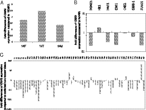
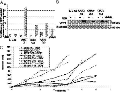
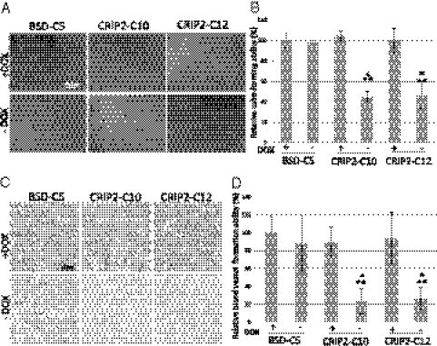
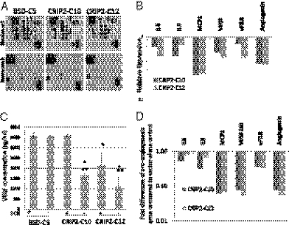
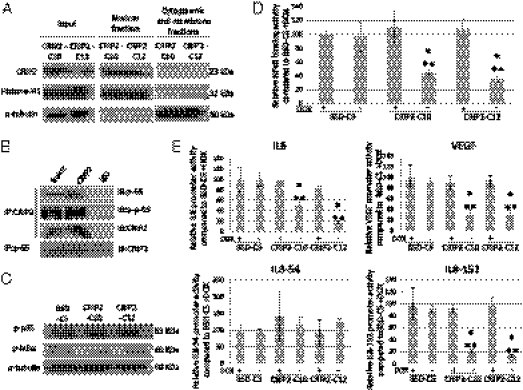
Chromosome 14 was transferred into tumorigenic nasopharyngeal carcinoma and esophageal carcinoma cell lines by a microcell-mediated chromosome transfer approach. Functional complementation of defects present in the cancer cells suppressed tumor formation. A candidate tumor-suppressor gene, cysteine-rich intestinal protein 2 (CRIP2), located in the hot spot for chromosomal loss at 14q32.3, was identified as an important candidate gene capable of functionally suppressing tumor formation. Previous studies have shown that CRIP2 is associated with development. To date, no report has provided functional evidence supporting a role for CRIP2 in tumor development. The present study provides unequivocal evidence that CRIP2 can functionally suppress tumorigenesis. CRIP2 is significantly down-regulated in nasopharyngeal carcinoma cell lines and tumors. CRIP2 reexpression functionally suppresses in vivo tumorigenesis and angiogenesis; these effects are induced by its transcription-repressor capability. It interacts with the NF-κB/p65 to inhibit its DNA-binding ability to the promoter regions of the major proangiogenesis cytokines critical for tumor progression, including IL6, IL8, and VEGF. In conclusion, we provide compelling evidence that CRIP2 acts as a transcription repressor of the NF-κB-mediated proangiogenic cytokine expression and thus functionally inhibits tumor formation and angiogenesis.


Conflict of interest statement

The authors declare no conflicts of interest.

Figures

Fig. 1.
Fig. 1.
CRIP2 gene expression analysis. (A) qPCR analysis of CRIP2 expression in three chromosome 14 MCHs. The expression fold changes were compared with the recipient HONE1 cell line. (B) qPCR analysis of CRIP2 expression in seven NPC cell lines. The expression fold changes of the NPC cell lines were compared with that of the immortalized nontumorigenic cell line NP460. (C) qPCR analysis of CRIP2 expression in 60 NPC paired biopsy specimens. The fold changes of the NPC tumor tissues were compared with their corresponding nontumor tissues.
Fig. 2.
Fig. 2.
CRIP2 expression levels in the CRIP2-expressing clones in an in vivo tumorigenicity assay. (A) qPCR analysis of CRIP2 expression in the vector-alone BSD-C5 (±dox) and the three CRIP2-expressing clones CRIP2-C9, -C10, and -C12 (±dox). The expression levels were compared with that of BSD-C5 (+dox). The NP460 cell line was used as a control for endogenous CRIP2 expression. (B) Western blot analysis of BSD-C5 and CRIP2-C9, -C10, and -C12 (±dox). α-tubulin served as a loading control. (C) In vivo tumorigenicity assay of CRIP2-expressing clones and vector-alone controls (+dox). The curves represent an average tumor volume of all sites inoculated for each cell population. Vector-alone BSD-C5 (±dox) formed large tumors by 6 wk postinjection. Small tumors were induced after 5–6 wk postinjection in all of the CRIP2-expressing clones. Tumorigenicity was restored when CRIP2 gene expression was switched off (+dox).
Fig. 3.
Fig. 3.
In vitro and in vivo angiogenesis assays of CRIP2-expressing clones. (A) Representative images of the HUVEC tube formation assay of vector-alone (BSD-C5) and CRIP2-expressing clones. (B) Summary of the relative tube-forming ability of the vector-alone (BSD-C5) and CRIP2-expressing clones (±dox). Relative tube-forming ability was calculated by comparing the total tube length of each sample to that of BSD-C5 (+dox). (C) Representative images of the in vivo Matrigel plug CD34 IHC staining of vector-alone (BSD-C5) and CRIP2-expressing clones. The brown color represents positive staining of the blood vessels. (D) Summary of the relative blood vessel formation ability of vector-alone (BSD-C5) and CRIP2-expressing clones (±dox). Relative blood vessel formation ability was calculated by comparing the total tube length of each sample with that of the BSD-C5 (+dox) control. *P < 0.05 and **P < 0.05, statistically significant differences compared with the vector-alone control and corresponding +dox control, respectively.
Fig. 4.
Fig. 4.
Angiogenesis-related protein expression in CRIP2-expressing clones. (A) Results of the angiogenesis protein array of the vector-alone BSD-C5 and CRIP2-expressing clones. Duplicate spots: 1, angiogenin; 2, IL-6; 3, IL-8; 4, MCP1; 5, PDGF-BB; 6, RANTES; 7, VEGF; 8, uPAR. (B) Summary of the relative expression of angiogenesis-related proteins in the CRIP2-expressing clones. Relative expression was calculated by comparing the intensity on the protein array of CRIP2-expressing clones vs. the vector-alone. (C) VEGF ELISA of the vector-alone and CRIP2-expressing clones. The total VEGF protein concentration (pg/mL) in the conditioned media of the vector-alone and CRIP2-expressing clones (+dox) was determined. (D) qPCR analysis verifying mRNA transcription levels of candidate genes identified by angiogenesis protein array in the CRIP2-expressing clones. The fold changes were compared with those of the vector-alone BSD-C5. *P < 0.05 and **P < 0.05, statistically significant differences compared with the vector-alone control and corresponding +dox control, respectively.
Fig. 5.
Fig. 5.
Transcriptional regulatory role of CRIP2. (A) Subcellular fractionation of CRIP2-expressing clones. Input protein, nuclear fraction, and cytoplasmic and membrane fraction were used for Western blot analysis. Anti–histone-1H and anti–α-tubulin antibodies were used as positive controls for nuclear and cytoplasmic and membrane fractions, respectively. (B) Co-IP assay using CRIP2 and NF-κB/p65 antibodies. CRIP2 and the total and phosphorylated NF-κB/p65 were detected in the CRIP2-expressing clones after co-IP. Anti-rabbit IgG was used as a negative control. (C) Phosphorylation status of p65 and IκBα in CRIP2-expressing clones. α-tubulin served as a loading control. (D) NF-κB–binding ability assay. The relative NF-κB–binding abilities of CRIP2-expressing clones were compared with those of the BSD-C5 (+dox) control. (E) Promoter assay results for IL-6, VEGF, and IL-8 promoters. Relative promoter activities were compared with the BSD-C5 (+dox) control. *P < 0.05 and **P < 0.05, statistically significant differences compared with the vector-alone control and corresponding +dox control, respectively.

References

    1. Cheung AK, et al. Chromosome 14 transfer and functional studies identify a candidate tumor-suppressor gene, Mirror image polydactyly 1, in nasopharyngeal carcinoma. Proc Natl Acad Sci USA. 2009;106:14478–14483. - PMC - PubMed
    1. Hui AB, et al. Detection of recurrent chromosomal gains and losses in primary nasopharyngeal carcinoma by comparative genomic hybridisation. Int J Cancer. 1999;82:498–503. - PubMed
    1. Lo KW, et al. High-resolution allelotype of microdissected primary nasopharyngeal carcinoma. Cancer Res. 2000;60:3348–3353. - PubMed
    1. Hoshi M, et al. Detailed deletion mapping of chromosome band 14q32 in human neuroblastoma defines a 1.1-Mb region of common allelic loss. Br J Cancer. 2000;82:1801–1807. - PMC - PubMed
    1. Ohta M, et al. Monocyte chemoattractant protein-1 expression correlates with macrophage infiltration and tumor vascularity in human esophageal squamous cell carcinomas. Int J Cancer. 2002;102:220–224. - PubMed

Publication types

MeSH terms

LinkOut - more resources