. 2011 May;121(5):1782-96.
doi: 10.1172/JCI45416. Epub 2011 Apr 11.

Protective T cell immunity in mice following protein-TLR7/8 agonist-conjugate immunization requires aggregation, type I IFN, and multiple DC subsets

Affiliations

Protective T cell immunity in mice following protein-TLR7/8 agonist-conjugate immunization requires aggregation, type I IFN, and multiple DC subsets

Kathrin Kastenmüller et al. J Clin Invest. 2011 May.

Abstract

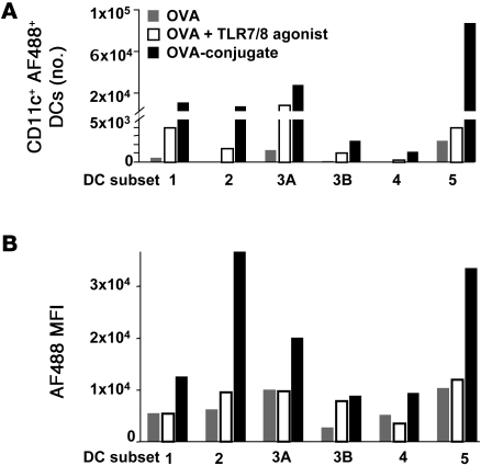
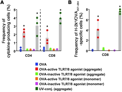
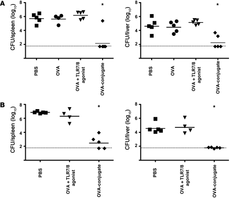
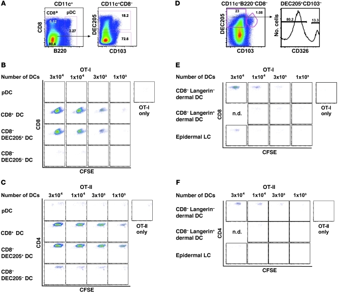
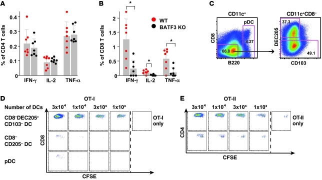
The success of a non-live vaccine requires improved formulation and adjuvant selection to generate robust T cell immunity following immunization. Here, using protein linked to a TLR7/8 agonist (conjugate vaccine), we investigated the functional properties of vaccine formulation, the cytokines, and the DC subsets required to induce protective multifunctional T cell immunity in vivo. The conjugate vaccine required aggregation of the protein to elicit potent Th1 CD4+ and CD8+ T cell responses. Remarkably, the conjugate vaccine, through aggregation of the protein and activation of TLR7 in vivo, led to an influx of migratory DCs to the LN and increased antigen uptake by several resident and migratory DC subsets, with the latter effect strongly influenced by vaccine-induced type I IFN. Ex vivo migratory CD8-DEC205+CD103-CD326- langerin-negative dermal DCs were as potent in cross-presenting antigen to naive CD8+ T cells as CD11c+CD8+ DCs. Moreover, these cells also influenced Th1 CD4+ T cell priming. In summary, we propose a model in which broad-based T cell-mediated responses upon vaccination can be maximized by codelivery of aggregated protein and TLR7/8 agonist, which together promote optimal antigen acquisition and presentation by multiple DC subsets in the context of critical proinflammatory cytokines.


Figures

Figure 1
Figure 1. OVA vaccines induce distinct changes in the composition of DCs within the DLN.
(A) Comparison of uptake of AF488-labeled OVA-conjugate (OVA-conj.) versus AF488-labeled OVA, with or without varying amounts of free TLR7/8 agonist 48 hours after immunization in Balb/c mice. Numbers within histograms refer to the percentages of CD11c+ DCs that took up the AF488-labeled OVA protein. Data are representative of 2 independent experiments. (B) Following enrichment of CD11c+ DCs from B6 mice (Supplemental Figure 1A), cells were divided into CD8+ DCs (subset 1) and pDCs (subset 2) based on their expression of CD8 and B220. B220CD8 DCs were further categorized as DEC205+CD103+ (subset 4), DEC205+CD103, and DEC205CD103 (subset 5) DC populations based on their expression of CD103 and DEC205, and DEC205+CD103 cells were further divided into CD326-positive (Epcam-positive) (subset 3B) and -negative populations (subset 3A). (C) Proportions of the different DC subpopulations in LNs of PBS-treated mice. Each DC subset was assigned a number and color based on the gating strategy depicted in B. Phenotypic descriptions of the subsets with the matching color codes are shown. The pie chart shows the relative percentage of each DC subset within the total CD11c+ DC population. Data were collected from pooled DLNs of 10 immunized mice. (D) Absolute numbers of cells in different DC subpopulations analyzed at different time points after immunization with either OVA, OVA plus free TLR7/8 agonist, or OVA-conjugate (n = 10 mice/group). The distribution of the CD11c+ DC subpopulations in the DLNs is presented as absolute number and the relative proportion (pie charts) of each DC subset at various times after immunization.
Figure 2
Figure 2. DC subsets differentially take up OVA vaccines.
Twenty-four hours after immunization of B6 mice with 20 μg AF488-labeled OVA, AF488-labeled OVA plus free TLR7/8 agonist, or AF488-labeled OVA-conjugate DLNs were harvested, pooled, and enriched for CD11c+ DCs. (A) Absolute numbers of the different DC subsets that are AF488 positive. (B) MFI of AF488-positive cells within each DC subset. DC subset 1, CD8+ DCs; DC subset 2, pDCs; DC subset 3A, langerin-negative dermal DCs; DC subset 3B, epidermal LCs; DC subset 4, langerin-positive dermal DCs; DC subset 5, CD8DEC205 (resident, blood-derived) DCs.
Figure 3
Figure 3. Protein aggregation and IFN-α enhance antigen uptake by CD11c+ DCs in vivo.
(A) Representative elution profiles of unconjugated OVA and UV-conjugated or chemically conjugated (SMCC-conj.) OVA, using fast protein liquid chromatography. (B) BALB/c mice (n = 10) were immunized with the eluted fraction (monomer or aggregate) of the different conjugates linked to AF488, and the percentage of CD11c+ DCs that took up AF488 was analyzed 24 hours later. (C) Uptake of OVA-conjugate was assessed in CD11c+ DCs isolated from the DLNs from B6, TLR7 KO, IFN-αβ receptor KO, and IL-12p40 KO mice. In some separate experiments, exogenous IFN-α was injected with the conjugate vaccine or anti–IFNαR-1 was administered intraperitoneally before immunization. (B and C) Numbers within histograms refer to the percentages of CD11c+ DCs that took up the AF488-labeled OVA protein. Data are representative of at least 2 independent experiments.
Figure 4
Figure 4. Protein aggregation is required for the induction of OVA-specific T cell responses.
B6 mice (n = 3/group) were immunized (s.c.) twice, 3 weeks apart, with 20 μg monomeric or aggregated OVA protein conjugated to an active or inactive TLR7/8 agonist or OVA protein alone. Seven days after the second immunization with the indicated conjugate vaccines, T cells in spleens were analyzed for cytokine production or tetramer binding by flow cytometry. (A) Frequency of OVA-specific IFN-γ–, IL-2–, or TNF-α–producing CD4+ or CD8+ T cells. (B) Frequency of H-2Kb/OVA257–264 tetramer–specific CD8+ T cells *P < 0.05, comparing aggregated protein conjugated with an active TLR7/8 agonist or the UV-conjugate compared with all other vaccine groups. Each symbol represents an individual mouse.
Figure 5
Figure 5. OVA-conjugate vaccine confers protection against L. monocytogenes challenge.
(A) Mice (n = 4-5) were immunized twice, 3 weeks apart, with PBS, 20 μg OVA, OVA plus free TLR7/8 agonist, or the OVA-conjugate vaccine. Fourteen days after the second immunization mice were challenged i.v. with 3 × 10⁴ CFUs of recombinant L. monocytogenes OVA. Bacterial loads in spleen and liver were enumerated 3 days after the challenge. (B) Mice (n = 4–5) were immunized once with 20 μg OVA plus free TLR7/8 agonist or the OVA-conjugate vaccine or were left untreated. Six weeks after immunization mice were challenged i.v. with 6 × 10⁴ CFUs of recombinant L. monocytogenes OVA. Bacterial loads in spleen and liver were enumerated 3 days after the challenge. Solid lines represent the geometrical mean. Dotted lines represents the level of detection. *P < 0.05, Mann-Whitney test, comparing OVA-conjugate with all other vaccine groups. Each symbol represents an individual mouse.
Figure 6
Figure 6. TLR7 and type I IFN influence T cell priming after immunization with OVA-conjugate vaccine.
B6, TLR7 KO, or IFN-αβ receptor (R) KO mice (n = 4/group) were immunized as described above (see Figure 5). As indicated for some groups, IFN-α was given together with the conjugate vaccine. (A) Frequency of OVA-specific IFN-γ–, IL-2–, or TNF-α–producing CD4+ or CD8+ T cells. (B) Frequency of H-2Kb/OVA257–264 tetramer–specific CD8+ T cells. Each symbol represents an individual mouse. Data are representative of 2 independent experiments. *P < 0.05; **P < 0.01.
Figure 7
Figure 7. Resident CD8+ DCs and migratory CD205+CD103 dermal DCs provide enhanced antigen presentation to naive CD4+ and CD8+ T cells.
B6 mice (n = 35–50) were immunized with 20 μg OVA-conjugate, and, 48 hours after immunization, DLNs were pooled, sorted, and cocultured with 5 × 10⁴ CFSE-labeled OT-I or OT-II T cells. (A and D) Gating strategy for sorting of DC subsets. Numbers in gates represent the frequency of gated cells. The purity of the cell populations assessed after the sort was approximately 96%. CFSE profiles of (B and E) OT-I or (C and F) OT-II cells cocultured for 60 hours with serial dilution of sorted DC subsets. OT-I (CD8) or OT-II (CD4) cells without DCs served as negative controls. Each stimulation condition was performed in duplicate (n.d., not done). Data are representative of 3 separate experiments.
Figure 8
Figure 8. CD8DEC205+CD103 dermal DCs influence Th1 CD4+ and CD8+ T cell immunity.
BATF3 KO mice or littermate controls (WT) were immunized with OVA-conjugate. Splenocytes were harvested 10 days after immunization, and antigen-specific cytokine production was analyzed for (A) CD4+ or (B) CD8+ T cells. Results shown represent the mean ± SD of 2 independent experiments with n = 3–4 mice per group. DEC205+CD103 dermal DCs efficiently induce CD4+ and CD8+ T cell proliferation. BATF3 KO mice (n = 17) were immunized with the OVA-conjugate vaccine, and DLNs were harvested and pooled 48 hours after immunization. DC subsets were immediately sorted ex vivo and cocultured with 5 × 10⁴ CFSE-labeled naive OT-I or OT-II T cells. (C) Gating strategy for sorting of DC subsets. Numbers in gates represent the frequency of gated cells. The purity of the cell populations assessed after the sort was more than 96%. CFSE profiles of (D) OT-I or (E) OT-II cells cocultured for 60 hours with serial dilution of sorted DC subsets. As negative controls, OT-I (CD8) or OT-II (CD4) cells alone were used. Each symbol represents an individual mouse. Each stimulation was performed in duplicate. *P < 0.05.

References

    1. Plotkin SA. Vaccines: correlates of vaccine-induced immunity. Clin Infect Dis. 2008;47(3):401–409. doi: 10.1086/589862. - DOI - PubMed
    1. Reed SG, Bertholet S, Coler RN, Friede M. New horizons in adjuvants for vaccine development. Trends Immunol. 2009;30(1):23–32. doi: 10.1016/j.it.2008.09.006. - DOI - PubMed
    1. Gamvrellis A, Leong D, Hanley JC, Xiang SD, Mottram P, Plebanski M. Vaccines that facilitate antigen entry into dendritic cells. Immunol Cell Biol. 2004;82(5):506–516. doi: 10.1111/j.0818-9641.2004.01271.x. - DOI - PubMed
    1. Obst R, van Santen HM, Mathis D, Benoist C. Antigen persistence is required throughout the expansion phase of a CD4(+) T cell response. J Exp Med. 2005;201(10):1555–1565. doi: 10.1084/jem.20042521. - DOI - PMC - PubMed
    1. Trinchieri G. Interleukin-12 and the regulation of innate resistance and adaptive immunity. Nat Rev Immunol. 2003;3(2):133–146. - PubMed

Publication types

MeSH terms