

Utility of 3-year torso computed tomography and head imaging in asymptomatic patients with high-risk melanoma
- PMID: 21540750
- PMCID: PMC3131441
- DOI: 10.1097/CMR.0b013e3283471086
Utility of 3-year torso computed tomography and head imaging in asymptomatic patients with high-risk melanoma
Abstract
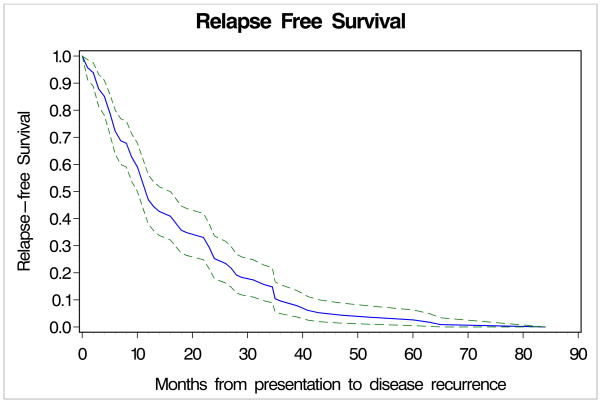
There is no general consensus regarding the optimal follow-up strategy for patients with melanoma. We sought to determine the utility and cost effectiveness of radiological restaging of patients with stage IIB-IIIC melanoma at the 3-year follow-up time point. A retrospective review of 210 patients diagnosed with stage IIB-IIIC melanoma seen in the Cutaneous Oncology Program at Beth Israel Deaconess Medical Center between January, 2001 and July, 2006 was conducted. Fifty-two patients were asymptomatic and continuously disease free and underwent restaging head computed tomography (CT) or MRI and torso CT scans 3 years after completion of local-regional therapy or initiation of adjuvant treatment. True positive, false positive and normal scans were identified and the cost per diagnosis calculated. Fifty-five percent of patients developed melanoma recurrences: 88% before 3 years (median time to recurrence 12 months, 95% confidence interval: 10-16 months). The majority of patients (69%) recurred with disease symptoms. Twenty-five head CT scans, 27 head MRIs, and 52 torso CTs were performed. One false-positive head CT and five abnormal torso CT scans (three false positive, two true positive) were identified. The total cost per diagnosis was $312,990. Extensive 3-year restaging imaging seems to be of limited value for symptomatic and continuously disease-free patients with stage IIB-IIIC melanoma. Furthermore, given the low risk of recurrence beyond 3 years, it is likely that subsequent routine imaging would have similarly low utility.
References
-
- National Cancer Institute. Surveillance Epidemiology and End Results. 2010.
-
- Balch CM, Soong SJ, Atkins MB, Buzaid AC, Cascinelli A, Coit DG, et al. An evidence-based staging system for cutaneous melanoma. CA Cancer J Clin. 2004;54:131–49. quiz 182-4. - PubMed
-
- Balch CM, Houghton A, Jr, Sober A, Soong SJ, Atkins MB, Thompson JF. Missouri: Quality Medical Publishing, Inc. 5. 2009. Cutaneous Melanoma; pp. 565–572.
-
- Aloia TA, Gershenwald JE, Andtbacka RH, Johnson MM, Schacherer CW, Ng CS, et al. Utility of computed tomography and magnetic resonance imaging staging before completion lymphadenectomy in patients with sentinel lymph node-positive melanoma. J Clin Oncol. 2006;24:2858–65. - PubMed
-
- Miranda EP, Gertner M, Wall J, Grace E, Kashani-Sabet M, Allen R, et al. Routine imaging of asymptomatic melanoma patients with metastasis to sentinel lymph nodes rarely identifies systemic disease. Arch Surg. 2004;139:831–6. discussion 836-7. - PubMed
Publication types
MeSH terms
Grants and funding
LinkOut - more resources
Full Text Sources
Medical
Research Materials
