. 2011 Jun;17(6):673-5.
doi: 10.1038/nm.2389. Epub 2011 May 3.

Identification of T helper type 1-like, Foxp3+ regulatory T cells in human autoimmune disease

Affiliations

Identification of T helper type 1-like, Foxp3+ regulatory T cells in human autoimmune disease

Margarita Dominguez-Villar et al. Nat Med. 2011 Jun.

Abstract

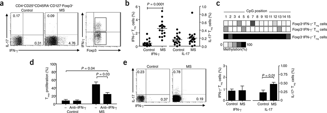
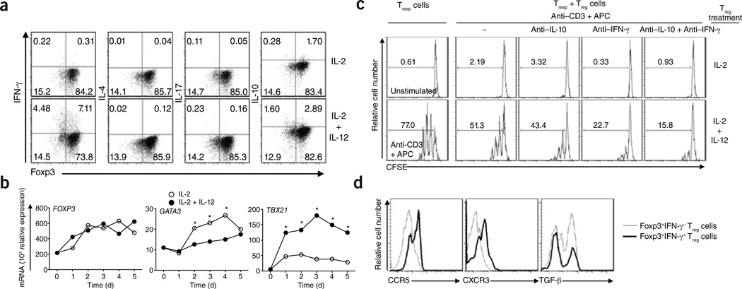
CD4(+)CD25(high)CD127(low/-) forkhead box p3 (Foxp3)(+) regulatory T cells (T(reg) cells) possess functional plasticity. Here we describe a higher frequency of T helper type 1 (T(H)1)-like, interferon-γ (IFN-γ)-secreting Foxp3(+) T cells in untreated subjects with relapsing remitting multiple sclerosis (RRMS) as compared to healthy control individuals. In subjects treated with IFN-β, the frequency of IFN-γ(+)Foxp3(+) T cells is similar to that in healthy control subjects. In vitro, human T(reg) cells from healthy subjects acquire a T(H)1-like phenotype when cultured in the presence of interleukin-12 (IL-12). T(H)1-like T(reg) cells show reduced suppressive activity in vitro, which can partially be reversed by IFN-γ-specific antibodies or by removal of IL-12.


Figures

Figure 1
Figure 1
Treg cells from individuals with RRMS secrete IFN-γ ex vivo. (a) The frequency of FACS-sorted IFN-γ+ and IL-17+ Treg cells in healthy control individuals (left) and untreated individuals with RRMS (middle, n = 17) gated on Foxp3+ Treg cells. Right, purity analysis of the sorted IFN-γ+Foxp3+ and IFN-γ Foxp3+ populations from subjects with RRMS used for methylation analysis in c. (b) Percentage of IFN-γ+Foxp3+ and IL-17+Foxp3+ Treg cells (n = 17) as a proportion of total Foxp3+ Treg cells. (c) Representative example of methylation analysis of the TSDR region of the FOXP3 locus in sorted IFN-γ+Foxp3+ and IFN-γ Foxp3+ Treg cells from subjects with RRMS. An analysis of IFN-γ+Foxp3 memory T cells from subjects with RRMS is shown as a control. (d) Proliferation of responder T (Tresp) cells cultured with ex vivo FACS-sorted Treg cells from healthy control subjects and untreated subjects with multiple sclerosis (MS; Treg cell:Tresp cell ratio of 1:2) in the presence or absence of an IFN-γ–specific antibody (n = 4). (e) The frequency of IFN-γ+ and IL-17+ Treg cells in healthy control subjects (left) or IFN-β–treated patients with RRMS (right) as assessed by intracellular cytokine staining and FACS analysis. The bar diagram (right) shows the percentage of IFN-γ+Foxp3+ and IL-17+Foxp3+ cells as a proportion of total Foxp3+ Treg cells in healthy controls or IFN-β–treated patients with RRMS (n = 12). Approval for studies was obtained from the Brigham and Women’s Hospital Institutional Review Board, and informed consent was obtained from all donors.
Figure 2
Figure 2
Characterization of IL-12–driven, IFN-γ+Foxp3+ Treg cells in vitro from healthy controls. (a) Intracellular staining for IFN-γ, IL-4, IL-17 and IL-10 of untreated (upper row) and IL-12-stimulated (bottom row) human Treg cells from healthy controls at day 4. (b) mRNA expression of FOXP3 (left), GATA3 (middle) and TBX21 (right) in Treg cells stimulated in the presence or absence of IL-12 for 5 d (data are a representative example of three experiments performed with similar results; *P < 0.05). (c) Proliferation of Tresp cells cocultured for 3 d with Treg cells (Treg cell:Tresp cell ratio of 1:2) previously treated with IL-2 (top) or IL-2 + IL-12 (bottom), as assessed by carboxyfluorescein succinimidyl ester (CFSE) dilution. Histograms depict unstimulated Tresp cells alone (top left), Tresp cells stimulated with antibody to CD3 (anti-CD3) and antigen-presenting cells without Treg cells (bottom left) and Tresp cell and Treg cell cocultures without blocking antibodies (second column), in the presence of an IL-10–specific blocking antibody (anti–IL-10; third column), IFN-γ–specific blocking antibody (anti–IFN-γ; fourth column) or both antibodies (fifth column). (d) Representative example of staining for TH1-associated chemokines(CCR5 and CXCR3) and TGF-β on IFN-γ+Foxp3+ and IFN-γ Foxp3+ Treg cells (data are representative of three experiments).

References

    1. McFarland HF, Martin R. Nat. Immunol. 2007;8:913–919. - PubMed
    1. Hafler DA, et al. N. Engl. J Med. 1985;312:1405–1411. - PubMed
    1. Viglietta V, Baecher-Allan C, Weiner HL, Hafler DA. J. Exp. Med. 2004;199:971–979. - PMC - PubMed
    1. Martinez-Forero I, et al. Eur. J. Immunol. 2008;38:576–586. - PubMed
    1. Kumar M, et al. J. Neuroimmunol. 2006;180:178–184. - PubMed

Publication types

MeSH terms