. 2010 May 26;1(5):69-80.
doi: 10.4331/wjbc.v1.i5.69.

Role of matricellular proteins in cardiac tissue remodeling after myocardial infarction

Affiliations

Role of matricellular proteins in cardiac tissue remodeling after myocardial infarction

Yutaka Matsui et al. World J Biol Chem. .

Abstract

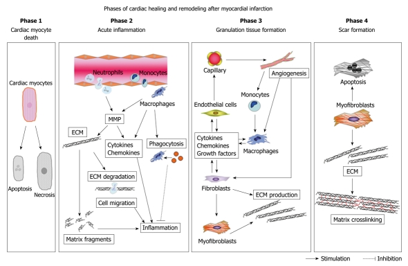
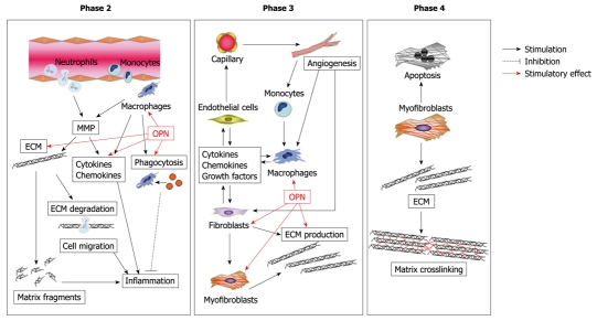
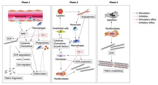
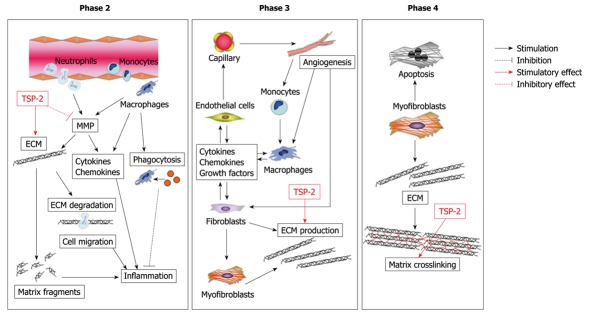
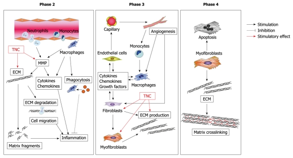
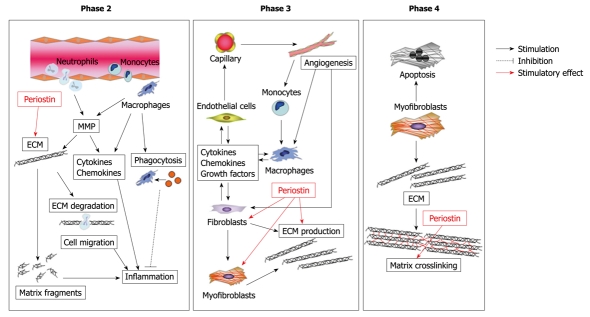
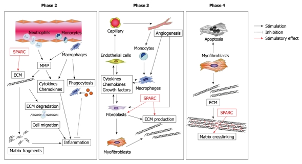
After onset of myocardial infarction (MI), the left ventricle (LV) undergoes a continuum of molecular, cellular, and extracellular responses that result in LV wall thinning, dilatation, and dysfunction. These dynamic changes in LV shape, size, and function are termed cardiac remodeling. If the cardiac healing after MI does not proceed properly, it could lead to cardiac rupture or maladaptive cardiac remodeling, such as further LV dilatation and dysfunction, and ultimately death. Although the precise molecular mechanisms in this cardiac healing process have not been fully elucidated, this process is strictly coordinated by the interaction of cells with their surrounding extracellular matrix (ECM) proteins. The components of ECM include basic structural proteins such as collagen, elastin and specialized proteins such as fibronectin, proteoglycans and matricellular proteins. Matricellular proteins are a class of non-structural and secreted proteins that probably exert regulatory functions through direct binding to cell surface receptors, other matrix proteins, and soluble extracellular factors such as growth factors and cytokines. This small group of proteins, which includes osteopontin, thrombospondin-1/2, tenascin, periostin, and secreted protein, acidic and rich in cysteine, shows a low level of expression in normal adult tissue, but is markedly upregulated during wound healing and tissue remodeling, including MI. In this review, we focus on the regulatory functions of matricellular proteins during cardiac tissue healing and remodeling after MI.

Keywords: Cardiac healing; Cardiac remodeling; Extracellular matrix; Matricellular proteins; Myocardial infarction.


Figures

Figure 1
Figure 1
Phases of cardiac healing and remodeling after myocardial infarction (MI). The cardiac healing and remodeling process after MI can be divided into four phases: (1) death of cardiomyocytes; (2) acute inflammation; (3) formation of granulation tissue; and (4) scar formation. Death of cardiomyocytes starts at approximately 1 h after coronary artery occlusion, and can either be the result of apoptosis or necrosis. During acute inflammation, the influx of inflammatory cells, including neutrophils and monocytes, for phagocytosis and removal of dead cardiomyocytes into the infarcted area and degradation of the extracellular matrix (ECM) by matrix metalloproteinase (MMP) takes place between 1 h and 4 d after MI. MMP also modulates inflammatory cytokine and chemokine activity. Generation of matrix fragments exerts potent inflammatory effects. Thereafter, formation of granulation tissue, characterized by the presence of fibroblasts, macrophages, myofibroblasts, new blood vessels, and ECM proteins, occurs in the infarcted heart between 2 and 14 d after MI. To rescue the loss of the shielding effects of the normal matrix, fibroblasts and myofibroblasts produce ECM. Finally, granulation tissue matures in the infarcted heart between 14 d and 2 mo after MI. The scar is characterized by a cross-linked, collagen-rich region that is induced by lysyl oxidase. In this phase, most infarct myofibroblasts undergo apoptosis and disappear. The time intervals for each phase are dependent on the species, as rodents exhibit an accelerated inflammatory and reparative response following MI as compared with large mammals.
Figure 2
Figure 2
Possible role of osteopontin (OPN) in cardiac healing and remodeling after MI. OPN modulates various functions of macrophages, such as migration, phagocytosis, and pro-inflammatory cytokine production. OPN also regulates proliferation and adhesion of fibroblasts. Moreover, OPN modulates myofibroblast differentiation and function, thereby promoting collagen synthesis and organization.
Figure 3
Figure 3
Possible role of thrombospondin-1 (TSP-1) in cardiac healing and remodeling after MI. Expression of TSP-1 works as a barrier that suppresses inflammatory chemokines and cytokines via its transforming growth factor-β1 (TGF-β1)-activating effect, thereby preventing extension of the inflammatory process into the non-infarcted region. In addition, TSP-1 could affect maturation of the infarct scar through the activation of TGF-β1 and inhibition of MMP activity.
Figure 4
Figure 4
Possible role of TSP-2 in cardiac healing and remodeling after MI. TSP-2 is a crucial regulator of the integrity of the cardiac ECM during maturation of the infarct scar, presumably via regulation of MMP activity, and subsequent deposition of a cross-linked collagen matrix.
Figure 5
Figure 5
Possible role of tenascin-C (TNC) in cardiac healing and remodeling after MI. TNC stimulates fibroblasts to produce matrix, thereby causing cardiac fibrosis and maladaptive remodeling after MI. Furthermore, TNC upregulates transcription and activity of MMP and promotes degradation of ECM.
Figure 6
Figure 6
Possible role of periostin in cardiac healing and remodeling after MI. Periostin regulates the integrity of the ECM through its ability to bind multiple ECM proteins, thereby affecting the structural integrity of the adult heart matrix. In addition, periostin promotes migration of fibroblasts via interaction with integrin.
Figure 7
Figure 7
Possible role of secreted protein, acidic and rich in cysteine (SPARC) in cardiac healing and remodeling after MI. SPARC regulates collagen maturation by direct interaction with collagen type I fibers and regulation of fibronectin matrix assembly. SPARC mediates TGF-β1 signaling, which is involved in wound healing and collagen production, thereby regulating cardiac healing and remodeling.

References

    1. Rosamond W, Flegal K, Friday G, Furie K, Go A, Greenlund K, Haase N, Ho M, Howard V, Kissela B, et al. Heart disease and stroke statistics--2007 update: a report from the American Heart Association Statistics Committee and Stroke Statistics Subcommittee. Circulation. 2007;115:e69–e171. - PubMed
    1. Matsumura S, Iwanaga S, Mochizuki S, Okamoto H, Ogawa S, Okada Y. Targeted deletion or pharmacological inhibition of MMP-2 prevents cardiac rupture after myocardial infarction in mice. J Clin Invest. 2005;115:599–609. - PMC - PubMed
    1. Cohn JN, Ferrari R, Sharpe N. Cardiac remodeling--concepts and clinical implications: a consensus paper from an international forum on cardiac remodeling. Behalf of an International Forum on Cardiac Remodeling. J Am Coll Cardiol. 2000;35:569–582. - PubMed
    1. Lindsey ML, Mann DL, Entman ML, Spinale FG. Extracellular matrix remodeling following myocardial injury. Ann Med. 2003;35:316–326. - PubMed
    1. McCurdy S, Baicu CF, Heymans S, Bradshaw AD. Cardiac extracellular matrix remodeling: fibrillar collagens and Secreted Protein Acidic and Rich in Cysteine (SPARC) J Mol Cell Cardiol. 2010;48:544–549. - PMC - PubMed