

Gains, losses and changes of function after gene duplication: study of the metallothionein family
- PMID: 21541013
- PMCID: PMC3081807
- DOI: 10.1371/journal.pone.0018487
Gains, losses and changes of function after gene duplication: study of the metallothionein family
Abstract
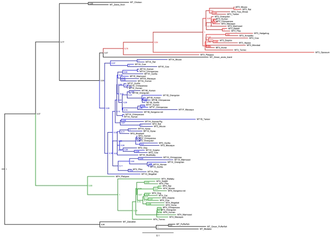
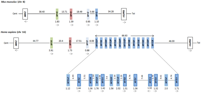
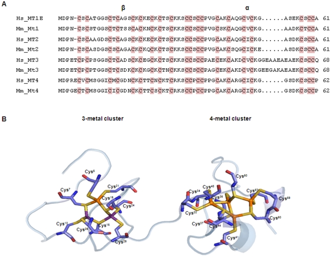
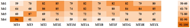
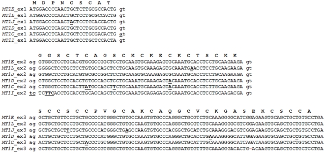
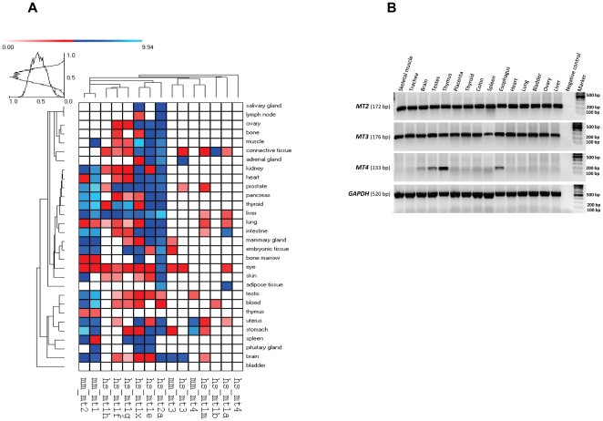
Metallothioneins (MT) are small proteins involved in heavy metal detoxification and protection against oxidative stress and cancer. The mammalian MT family originated through a series of duplication events which generated four major genes (MT1 to MT4). MT1 and MT2 encode for ubiquitous proteins, while MT3 and MT4 evolved to accomplish specific roles in brain and epithelium, respectively. Herein, phylogenetic, transcriptional and polymorphic analyses are carried out to expose gains, losses and diversification of functions that characterize the evolutionary history of the MT family. The phylogenetic analyses show that all four major genes originated through a single duplication event prior to the radiation of mammals. Further expansion of the MT1 gene has occurred in the primate lineage reaching in humans a total of 13 paralogs, five of which are pseudogenes. In humans, the reading frame of all five MT1 pseudogenes is reconstructed by sequence homology with a functional duplicate revealing that loss of invariant cysteines is the most frequent event accounting for pseudogeneisation. Expression analyses based on EST counts and RT-PCR experiments show that, as for MT1 and MT2, human MT3 is also ubiquitously expressed while MT4 transcripts are present in brain, testes, esophagus and mainly in thymus. Polymorphic variation reveals two deleterious mutations (Cys30Tyr and Arg31Trp) in MT4 with frequencies reaching about 30% in African and Asian populations suggesting the gene is inactive in some individuals and physiological compensation for its loss must arise from a functional equivalent. Altogether our findings provide novel data on the evolution and diversification of MT gene duplicates, a valuable resource for understanding the vast set of biological processes in which these proteins are involved.
Conflict of interest statement
Figures
Similar articles
-
Molecular evolution and functional divergence of the metallothionein gene family in vertebrates.J Mol Evol. 2014 Apr;78(3-4):217-33. doi: 10.1007/s00239-014-9612-5. Epub 2014 Feb 21. J Mol Evol. 2014. PMID: 24557429
-
Mammalian MT1 and MT2 metallothioneins differ in their metal binding abilities.Metallomics. 2013 Oct;5(10):1397-410. doi: 10.1039/c3mt00123g. Metallomics. 2013. PMID: 23925449
-
Molecular characterization of two metallothionein isoforms in avian species: evolutionary history, tissue distribution profile, and expression associated with metal accumulation.Comp Biochem Physiol C Toxicol Pharmacol. 2007 Apr;145(3):295-305. doi: 10.1016/j.cbpc.2006.10.012. Epub 2007 Jan 24. Comp Biochem Physiol C Toxicol Pharmacol. 2007. PMID: 17317328
-
Metallothionein polymorphisms in pathological processes.Metallomics. 2014 Jan;6(1):55-68. doi: 10.1039/c3mt00132f. Metallomics. 2014. PMID: 24068159 Review.
-
Human metallothionein expression under normal and pathological conditions: mechanisms of gene regulation based on in silico promoter analysis.Crit Rev Eukaryot Gene Expr. 2009;19(4):301-17. doi: 10.1615/critreveukargeneexpr.v19.i4.40. Crit Rev Eukaryot Gene Expr. 2009. PMID: 19817707 Review.
Cited by
-
Barley metallothioneins: MT3 and MT4 are localized in the grain aleurone layer and show differential zinc binding.Plant Physiol. 2012 Jul;159(3):1125-37. doi: 10.1104/pp.112.197798. Epub 2012 May 11. Plant Physiol. 2012. PMID: 22582132 Free PMC article.
-
Exposure to Dichlorodiphenyldichloroethylene (DDE) and Metallothionein Levels in Rats Fed with Normocaloric or High-Fat Diet: A Review.Int J Mol Sci. 2020 Mar 10;21(5):1903. doi: 10.3390/ijms21051903. Int J Mol Sci. 2020. PMID: 32164371 Free PMC article. Review.
-
Oxidative pathways of apo, partially, and fully Zn(II)- and Cd(II)-metalated human metallothionein-3 are dominated by disulfide bond formation.FEBS J. 2025 Feb;292(3):619-634. doi: 10.1111/febs.17333. Epub 2024 Dec 1. FEBS J. 2025. PMID: 39617990 Free PMC article.
-
The Effects of Essential and Non-Essential Metal Toxicity in the Drosophila melanogaster Insect Model: A Review.Toxics. 2021 Oct 16;9(10):269. doi: 10.3390/toxics9100269. Toxics. 2021. PMID: 34678965 Free PMC article. Review.
-
Sost and its paralog Sostdc1 coordinate digit number in a Gli3-dependent manner.Dev Biol. 2013 Nov 1;383(1):90-105. doi: 10.1016/j.ydbio.2013.08.015. Epub 2013 Aug 29. Dev Biol. 2013. PMID: 23994639 Free PMC article.
References
-
- Hughes AL. Adaptive evolution after gene duplication. Trends Genet. 2002;18:433–434. - PubMed
-
- Lynch M, Katju V. The altered evolutionary trajectories of gene duplicates. Trends Genet. 2004;20:544–549. - PubMed
-
- Lynch M. The origins of genome architecture. Sunderland: Sinauer Associates; 2007. 389
-
- Ohno S. Evolution by gene duplication. Heidelberg: Springer-Verlag; 1970. 160
-
- Ohta T. How gene families evolve. Theor Popul Biol. 1990;37:213–219. - PubMed
Publication types
MeSH terms
Substances
LinkOut - more resources
Full Text Sources
Other Literature Sources
Research Materials
