. 2011:2011:506171.
doi: 10.1155/2011/506171. Epub 2011 Mar 30.

Tocotrienol-rich fraction prevents cell cycle arrest and elongates telomere length in senescent human diploid fibroblasts

Affiliations

Tocotrienol-rich fraction prevents cell cycle arrest and elongates telomere length in senescent human diploid fibroblasts

Suzana Makpol et al. J Biomed Biotechnol. 2011.

Abstract

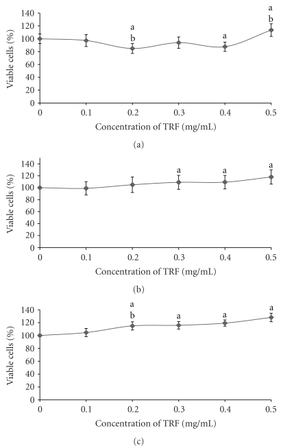
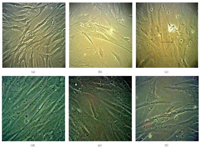
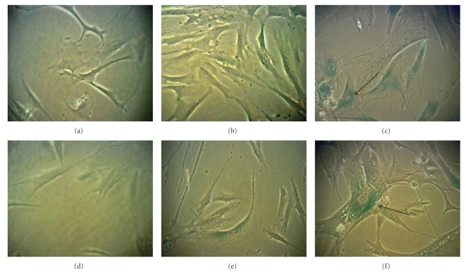
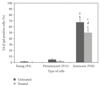
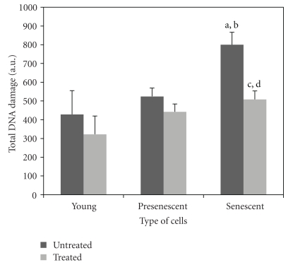
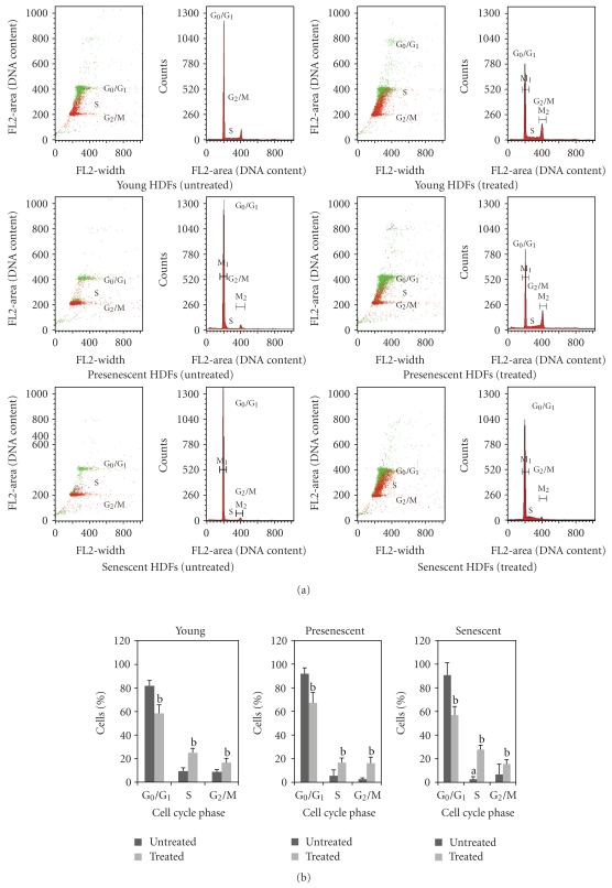
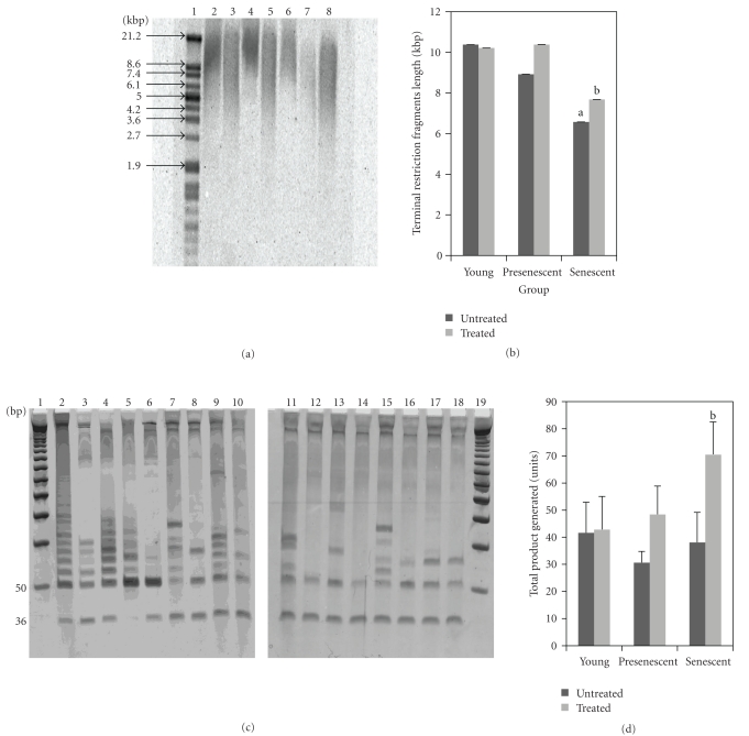
This study determined the molecular mechanisms of tocotrienol-rich fraction (TRF) in preventing cellular senescence of human diploid fibroblasts (HDFs). Primary culture of HDFs at various passages were incubated with 0.5 mg/mL TRF for 24 h. Telomere shortening with decreased telomerase activity was observed in senescent HDFs while the levels of damaged DNA and number of cells in G(0)/G(1) phase were increased and S phase cells were decreased. Incubation with TRF reversed the morphology of senescent HDFs to resemble that of young cells with decreased activity of SA-β-gal, damaged DNA, and cells in G(0)/G(1) phase while cells in the S phase were increased. Elongated telomere length and restoration of telomerase activity were observed in TRF-treated senescent HDFs. These findings confirmed the ability of tocotrienol-rich fraction in preventing HDFs cellular ageing by restoring telomere length and telomerase activity, reducing damaged DNA, and reversing cell cycle arrest associated with senescence.


Figures

Figure 1
Figure 1
Dose response of tocotrienol-rich fraction (TRF) on young (a), presenescent (b), and senescent (c) HDFs after 24 h incubation at 37°C. Incubation with TRF caused a significant increase in the viability of HDFs. aDenotes P < .05 compared to control, bP < .05 compared to lower concentration. Data are presented as mean ± SD, n = 9.
Figure 2
Figure 2
Morphological changes of HDFs in culture. Young (a), presenescent (b), and senescent (c) HDFs compared to TRF-treated HDFs (d)–(f). Senescent HDFs lost their original fibroblastic shape by acquiring a flattened morphology (indicated by arrow) with increased in size of nucleus and cells. The morphology of TRF-treated HDFs resembled that of young cells with more spindle-shaped cells present. Micrographs are shown at 200x magnification.
Figure 3
Figure 3
β-Galactosidase staining in young (a), presenescent (b) and senescent HDFs (c) compared to TRF-treated HDFs (d)–(f). Positive blue stain of SA-galactosidase appeared in senescent HDFs as indicated by arrow. Micrographs are shown at 200x magnification.
Figure 4
Figure 4
Quantitative analysis of positive β-galactosidase stained cells in HDFs during cellular ageing. The percentage of cells positive for SA-β-gal staining was significantly increased in senescent cells. Incubation of senescent cells with TRF significantly decreased the percentage of cells positive for SA-β-gal staining. aDenotes P < .05 compared to untreated young HDFs, and bP < .05 compared to untreated presenescent HDFs, cP < .05 compared to untreated senescent HDFs, dP < .05 compared to treated young HDFs, eP < .05 compared to treated presenescent HDFs. Data are presented as mean ± SEM, n = 6.
Figure 5
Figure 5
Comparison of total DNA damage at various stages of cellular ageing measured by Comet assay. Damaged DNA was higher in senescent HDFs which was decreased with TRF treatment. aDenotes P < .05 compared to untreated young HDFs, bP < .05 compared to untreated presenescent HDFs, cP < .05 compared to untreated senescent HDFs, dP < .05 compared to TRF-treated young HDFs. Data are presented as mean ± SD, n = 6.
Figure 6
Figure 6
Analysis of cell cycle progression. Flow cytometric analysis of cell cycle progression in young, presenescent, and senescent HDFs (a). Quantitative analysis of cell cycle progression in untreated and TRF-treated HDFs at various stages of cellular ageing (b). Cell population in the S phase was lower in senescent HDFs. Treatment with TRF significantly increased cells in S phase and G2/M phase for all stages of cellular ageing of HDFs. In contrast, cell populations in G0/G1 phase decreased significantly with TRF treatment. aDenotes P < .05 compared to S phase of untreated young HDFs, bP < .05 compared to untreated HDFs. Data are presented as mean ± SD, n = 6.
Figure 7
Figure 7
Effects of tocotrienol-rich fraction (TRF) on telomere length and telomerase activity. Representative Southern blot analysis of young, presenescent and senescent HDFs. Telomeric DNA is shown as wide smears in all lanes. Lane 1: molecular weight marker, lane 2: positive control DNA, lane 3: untreated young HDFs, lane 4: TRF-treated young HDFs, lane 5: untreated presenescent HDFs, lane 6: TRF-treated presenescent HDFs, lane 7: untreated senescent HDFs, lane 8: TRF-treated senescent HDFs (a). Telomere length (Terminal Restriction Fragments) of young, presenescent and senescent HDFs. Shortening of telomere length was observed with senescence of HDFs. Protective effects of TRF against telomere shortening was observed in senescent HDFs. aDenotes P < .05 compared to untreated young HDFs, bP < .05 compared to untreated senescent HDFs. Data are presented as mean ± SEM, n = 6 (b). Representative PCR analysis for telomerase activity of young, presenescent and senescent HDFs. Lane 1: molecular weight marker, lane 2: positive control, lane 3: positive control (heat treated), lane 4: TSR8 (1 μL), lane 5: TSR8 (2 μL), lane 6: negative control, lane 7: untreated young HDFs, lane 8: untreated young HDFs (heat treated), lane 9: TRF-treated young HDFs, lane 10: TRF-treated young HDFs (heat treated), lane 11: untreated presenescent HDFs, lane 12: untreated presenescent HDFs (heat treated), lane 13: TRF-treated presenescent HDFs, lane 14: TRF-treated presenescent HDFs (heat treated), lane 15: untreated senescent HDFs, lane 16: untreated senescent HDFs (heat treated), lane 17: TRF-treated senescent HDFs, lane 18: TRF-treated senescent HDFs (heat treated), lane 19: molecular weight marker (c). Telomerase activity (Total Product Generated, TPG) of young, presenescent and senescent HDFs. Treatment with TRF significantly increased the telomerase activity in senescent HDFs. aDenotes P < .05 compared to untreated senescent HDFs. Data is presented as mean ± SEM, n = 6 (d).

Similar articles

Cited by

References

    1. Hayflick L, Moorehead PS. The serial cultivation of human diploid cell strains. Experimental Cell Research. 1961;25(3):585–621. - PubMed
    1. Yeo EJ, Hwang YC, Kang CM, et al. Senescence-like changes induced by hydroxyurea in human diploid fibroblasts. Experimental Gerontology. 2000;35(5):553–571. - PubMed
    1. Campisi J. Cancer, aging and cellular senescence. In Vivo. 2000;14(1):183–188. - PubMed
    1. Duan J, Duan J, Zhang Z, Tong T. Irreversible cellular senescence induced by prolonged exposure to H2O2 involves DNA-damage-and-repair genes and telomere shortening. International Journal of Biochemistry and Cell Biology. 2005;37(7):1407–1420. - PubMed
    1. Finkel T. Oxidant signals and oxidative stress. Current Opinion in Cell Biology. 2003;15(2):247–254. - PubMed

Publication types

LinkOut - more resources