

A new case of spontaneous regression of inflammatory hepatic pseudotumor
- PMID: 21541218
- PMCID: PMC3085331
- DOI: 10.1155/2011/139125
A new case of spontaneous regression of inflammatory hepatic pseudotumor
Abstract
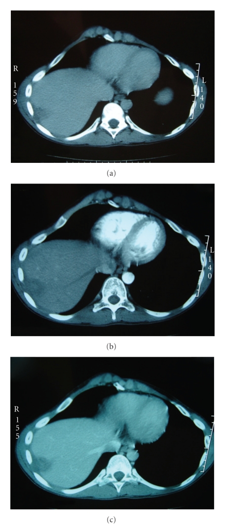
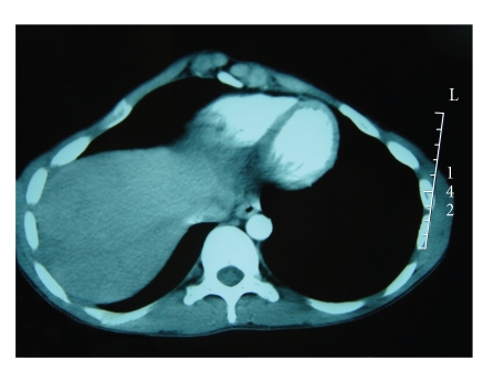
Introduction. Inflammatory pseudo-tumors (IPT) of the liver are rare and difficult to diagnose, because mimicking malignant tumors. Aim. We report a case of IPT of the liver wich diagnosis was made on clinical, radiological and evolutif features. Observation. A 15-year-old man had a 4-month history of abdominal pain in the right upper quadrant with fever and cought. Two successives ultrasonographies revealed a hypoechoic lesion occuping the segment VIII with 8 cm of diametre. Physical examination was normal. Laboratory investigation showed normal blood counts, liver function test and tumoral markers. Another ultrasonography was interpretated as normal. Tomodensitometry had showon a 3-cm lesion wich enhanced later after contrast injection. A second tomodensitometry done one mounth later described a 2-cm sub capsular heaptic lesion. Discussion. On routine activiy, pre operative diagnosis of IPT of the liver is difficut, and rarely made with certitude because mimicking a malignant tumor. In our cae report here, the analysis of previous history, of clinical, biological and radiological presentation, had permittes us to pose the diagnosis of PTI of the liver and this despite the absence of histological confirmation by percutaneous biopsy.
Figures


References
-
- Ishak KG, Anthony PP, Sobin LH. Histologic Typing of Tumors of the Liver. World Health Organisation: International Histologic Classification of Tumors. 2nd edition. Berlin, Germany: Springer; 1994.
-
- Koea JB, Broadhurst GW, Rodgers MS, McCall JL. Inflammatory pseudotumor of the liver: demographics, diagnosis, and the case for nonoperative management. Journal of the American College of Surgeons. 2003;196(2):226–235. - PubMed
-
- Horiuchi R, Uchida T, Kojima T, Shikata T. Inflammatory pseudotumor of the liver. Clinicopathologic study and review of the literature. Cancer. 1990;65(7):1583–1590. - PubMed
-
- Abbey-Toby A, Cazals-Hatem D, Colombat M, Belghiti J, Vilgrain V, Degott C. Inflammatory pseudo-tumor of the liver: is pre-operative diagnosis possible? Gastroenterologie Clinique et Biologique. 2003;27(10):883–890. - PubMed
-
- Yamaguchi J, Sakamoto Y, Sano T, Shimada K, Kosuge T. Spontaneous regression of inflammatory pseudotumor of the liver: report of three cases. Surgery Today. 2007;37(6):525–529. - PubMed
Publication types
LinkOut - more resources
Full Text Sources
