. 2011 Apr 26;6(4):e19274.
doi: 10.1371/journal.pone.0019274.

Expression of a dominant negative CELF protein in vivo leads to altered muscle organization, fiber size, and subtype

Affiliations

Expression of a dominant negative CELF protein in vivo leads to altered muscle organization, fiber size, and subtype

Dara S Berger et al. PLoS One. .

Abstract

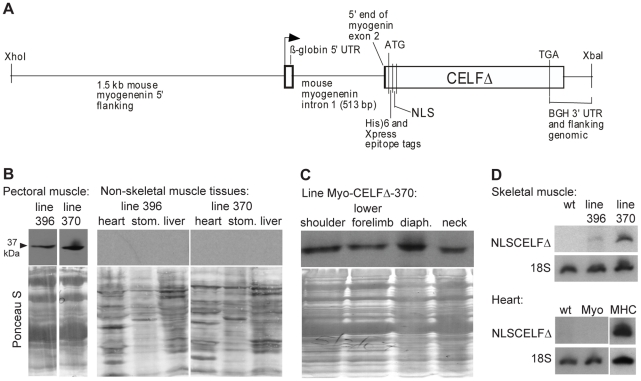
Background: CUG-BP and ETR-3-like factor (CELF) proteins regulate tissue- and developmental stage-specific alternative splicing in striated muscle. We previously demonstrated that heart muscle-specific expression of a nuclear dominant negative CELF protein in transgenic mice (MHC-CELFΔ) effectively disrupts endogenous CELF activity in the heart in vivo, resulting in impaired cardiac function. In this study, transgenic mice that express the dominant negative protein under a skeletal muscle-specific promoter (Myo-CELFΔ) were generated to investigate the role of CELF-mediated alternative splicing programs in normal skeletal muscle.

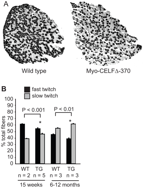
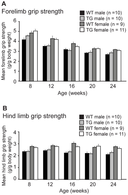
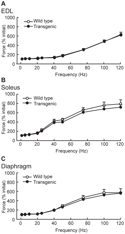
Methodology/principal findings: Myo-CELFΔ mice exhibit modest changes in CELF-mediated alternative splicing in skeletal muscle, accompanied by a reduction of endomysial and perimysial spaces, an increase in fiber size variability, and an increase in slow twitch muscle fibers. Weight gain and mean body weight, total number of muscle fibers, and overall muscle strength were not affected.

Conclusions/significance: Although these findings demonstrate that CELF activity contributes to the normal alternative splicing of a subset of muscle transcripts in vivo, the mildness of the effects in Myo-CELFΔ muscles compared to those in MHC-CELFΔ hearts suggests CELF activity may be less determinative for alternative splicing in skeletal muscle than in heart muscle. Nonetheless, even these small changes in CELF-mediated splicing regulation were sufficient to alter muscle organization and muscle fiber properties affected in myotonic dystrophy. This lends further evidence to the hypothesis that dysregulation of CELF-mediated alternative splicing programs may be responsible for the disruption of these properties during muscle pathogenesis.


Conflict of interest statement

Competing Interests: The authors have declared that no competing interests exist.

Figures

Figure 1
Figure 1. Myo-CELFΔ transgenic mice express the nuclear dominant negative CELF protein in skeletal muscles.
(A) The Myo-CELFΔ transgene contains NLSCELFΔ behind the mouse myogenin promoter. (B) Expression of NLSCELFΔ protein in skeletal muscle, but not heart, stomach (stom.), or liver, was confirmed in total protein lysates from tissues of Myo-CELFΔ mice by western blot using an antibody against the N-terminal Xpress epitope tag. Equivalent loading was confirmed by Ponceau S staining. Intervening lanes with samples from non-expressing lines were excised from the skeletal muscle blot. (C) Expression of NLSCELFΔ protein in a variety of Myo-CELFΔ skeletal muscles was confirmed by western blot using an antibody against the N-terminal Xpress epitope tag. Protein loading and integrity was confirmed by Ponceau S staining. (D) Expression levels of NLSCELFΔ were compared between Myo-CELFΔ lines by semi-quantitative RT-PCR. Lack of NLSCELFΔ transcripts in the Myo-CELFΔ heart (Myo) was confirmed; RNA from the heart of an MHC-CELFΔ-10 mouse (MHC), in which NLSCELFΔ is highly expressed in cardiac muscle, was used as a positive control.
Figure 2
Figure 2. The alternative splicing of some transcripts is affected in Myo-CELFΔ muscle.
The alternative splicing of transcripts that were previously identified as targets of CELF regulation in the mouse heart was evaluated in the pectoral muscles of nonparous female wild type and Myo-CELFΔ-370 siblings at 24 weeks. H2afy mutually exclusive exons 6 and 7 (A) and Capzb exon 8 (B) exhibited significant changes in splicing. (C) C10orf97 exon 5 showed a trend towards decreased inclusion in pectoral muscle. Although not statistically significant in pectoral muscle, a significant decrease in exon inclusion was observed in thigh muscle (data not shown; see text). (D) Nrap exon 12, which is repressed in DM1 skeletal muscle, displayed a significant increase in inclusion in Myo-CELFΔ muscle. An asterisk denotes a significant difference from wild type (P≤0.05).
Figure 3
Figure 3. Myo-CELFΔ mice have altered skeletal muscle organization and variable muscle fiber size.
(A) Transverse cross-sections of the lower forelimb of adult Myo-CELFΔ-370 mice reveal a reduction in the endomysial and perimysial spaces relative to that of a wild type littermate. Sections shown are representative of three independently processed sex- and age-matched littermate pairs. Higher magnification views of muscles from wild type (B) and Myo-CELFΔ-370 (C) littermates or wild type (D) and Myo-CELFΔ-396 (E) littermates reveal an increased prevalence of both enlarged (arrows) and diminutive (arrowheads) muscle fibers in transgenic mice. In order to ensure that the same muscle groups are being compared between wild type and transgenic mice, the regions shown in (B) and (C) are from a portion of the cross-section where the reduction in spacing around the muscle fibers in the Myo-CELFΔ-370 limb was less pronounced. (F) Fiber area was measured for muscle fibers within matched regions of hind limb sections from two sex- and age-matched MyoCELFΔ-370 and wild type pairs. Fiber size varied more in transgenic muscles, with an enrichment of both smaller (arrowheads) and larger (arrows) fibers relative to wild type muscles.
Figure 4
Figure 4. Fiber type composition is altered in Myo-CELFΔ mice.
(A) Representative sections of soleus muscles isolated from 15 week old wild type and Myo-CELFΔ-370 littermates that have been stained for myofibrillar actomyosin ATPase activity are shown. Pale fibers  =  slow twitch (Type I), dark fibers  =  fast twitch (Type II). (B) The number of slow twitch (pale) and fast twitch (dark) fibers were counted in 15 week old or 6–12 month old wild type and Myo-CELFΔ-370 littermates and mean values were calculated for each group. An asterisk denotes a significant difference from wild type (P≤0.05).
Figure 5
Figure 5. Grip strength is unaffected in Myo-CELFΔ mice.
Forelimb (A) and hind limb (B) grip strengths were measured every four weeks in a cohort of wild type and Myo-CELFΔ-370 siblings over a sixteen week time course. Force measurements were normalized against body weight. Males and females were assessed independently due to sex differences in muscle mass and body weight. No significant differences were found between sex-matched wild type and transgenic mice at any age evaluated.
Figure 6
Figure 6. Force measurements are not affected in skeletal muscles from Myo-CELFΔ mice.
Force was assessed ex vivo in extensor digitorum longus (EDL) (A), soleus (B), and diaphragm (C) muscles isolated from sex- and age-matched wild type and Myo-CELFΔ-370 mice. No significant differences were found at any frequency.

Similar articles

Cited by

References

    1. Pan Q, Shai O, Lee LJ, Frey BJ, Blencowe BJ. Deep surveying of alternative splicing complexity in the human transcriptome by high-throughput sequencing. Nat Genet. 2008;40(12):1413–1415. - PubMed
    1. Wang ET, Sandberg R, Luo S, Khrebtukova I, Zhang L, et al. Alternative isoform regulation in human tissue transcriptomes. Nature. 2008;456(7221):470–476. - PMC - PubMed
    1. Charlet-B N, Logan P, Singh G, Cooper T. Dynamic antagonism between ETR-3 and PTB regulates cell type-specific alternative splicing. Mol Cell. 2002;9:649–658. - PubMed
    1. Kalsotra A, Xiao X, Ward AJ, Castle JC, Johnson JM, et al. A postnatal switch of CELF and MBNL1 proteins reprograms alternative splicing in the developing heart. Proc Natl Acad Sci U S A. 2008;105(51):20333–20338. - PMC - PubMed
    1. Ladd A, Charlet-B N, Cooper T. The CELF family of RNA binding proteins is implicated in cell-specific and developmentally regulated alternative splicing. Mol Cell Biol. 2001;21(4):1285–1296. - PMC - PubMed

Publication types