. 2011 Apr 26;6(4):e19285.
doi: 10.1371/journal.pone.0019285.

Differential roles for STIM1 and STIM2 in store-operated calcium entry in rat neurons

Affiliations

Differential roles for STIM1 and STIM2 in store-operated calcium entry in rat neurons

Joanna Gruszczynska-Biegala et al. PLoS One. .

Abstract

The interaction between Ca(2+) sensors STIM1 and STIM2 and Ca(2+) channel-forming protein ORAI1 is a crucial element of store-operated calcium entry (SOCE) in non-excitable cells. However, the molecular mechanism of SOCE in neurons remains unclear. We addressed this issue by establishing the presence and function of STIM proteins. Real-time polymerase chain reaction from cortical neurons showed that these cells contain significant amounts of Stim1 and Stim2 mRNA. Thapsigargin (TG) treatment increased the amount of both endogenous STIM proteins in neuronal membrane fractions. The number of YFP-STIM1/ORAI1 and YFP-STIM2/ORAI1 complexes was also enhanced by such treatment. The differences observed in the number of STIM1 and STIM2 complexes under SOCE conditions and the differential sensitivity to SOCE inhibitors suggest their distinct roles. Endoplasmic reticulum (ER) store depletion by TG enhanced intracellular Ca(2+) levels in loaded with Fura-2 neurons transfected with YFP-STIM1 and ORAI1, but not with YFP-STIM2 and ORAI1, which correlated well with the number of complexes formed. Moreover, the SOCE inhibitors ML-9 and 2-APB reduced Ca(2+) influx in neurons expressing YFP-STIM1/ORAI1 but produced no effect in cells transfected with YFP-STIM2/ORAI1. Moreover, in neurons transfected with YFP-STIM2/ORAI1, the increase in constitutive calcium entry was greater than with YFP-STIM1/ORAI1. Our data indicate that both STIM proteins are involved in calcium homeostasis in neurons. STIM1 mainly activates SOCE, whereas STIM2 regulates resting Ca(2+) levels in the ER and Ca(2+) leakage with the additional involvement of STIM1.


Conflict of interest statement

Competing Interests: The authors have declared that no competing interests exist.

Figures

Figure 1
Figure 1. Real-time PCR analysis of Stim1 and Stim2 mRNA levels in neurons.
mRNA was isolated from primary cultures of neurons and astrocytes. TaqMan primers and probes were used to quantify specific mRNA levels. (A) The Gfap astrocytic marker and Map2 neuronal marker are differentially expressed in cultures of cortical neurons (Cx), hippocampal neurons (Hip) and astrocytes (Astro), confirming the purity of the cultures. The expression was related to the Gapdh level set as 1 for every culture, using the 2−dCT formula (dCT = CTtarget − CTGapdh; CT is the cycle threshold). (B) Stim1 and Stim2 are expressed in neurons of the cortex (Cx) and hippocampus (Hip) and in cortical astrocytes (Astro). The expression was related to Gapdh as above. (C) The actual number of Stim1 and Stim2 mRNA molecules in 20 ng of RNA isolated from cortical neurons was quantified using a standard curve with serial dilutions of cloned Stim1 and Stim2. (D) The actual number of Stim1 and Stim2 mRNA molecules in laser-dissected hippocampal neurons was quantified as above.
Figure 2
Figure 2. Expression and distribution of endogenous STIM proteins in subcellular neuronal fractions.
(A) Immunoblots of selected marker proteins demonstrate high separation efficiency for subcellular compartments of neurons on cytosolic (C) and membrane (M) fractions. Proteins were analyzed using the anti-pan Cadherin (plasma membrane), anti-STIM2, anti-Calnexin (ER membrane), anti-STIM1, anti-actin (cytosolic), anti-GAPDH (cytosolic), and anti-ORAI1 antibodies. Notice that STIM and ORAI1 proteins are present only in the membrane fraction. (B) Neurons were incubated for 10 min with 2 mM Ca2+ (left, −/−), 2 µM TG in 0.5 mM EGTA (middle, +/−), or 2 µM TG in 0.5 mM EGTA followed by the addition of Ca2+ in the presence of ML9 for 5 min (right, +/+). The cells were then subjected to fractionation into cytosolic and membrane fractions, and proteins were analyzed by immunoblotting using anti-STIM1, anti-STIM2, anti-ORAI1, anti-p-Cadherin and anti-actin (loading controls) antibodies. Blots were developed using chemiluminescence. Only membrane subfractions are shown. The image shows the results from one representative experiment. Bars indicate mean ± SD from at least three separate experiments. STIM1 or STIM2 bands were normalized to the level of loading control p-Cadherin for each immunoblot. (C) Effect of ML9 on SOCE of nontransfected neurons after restoration of extracellular Ca2+. ML9 was added 5 min after the readdition of 2 mM CaCl2. Raw data are shown. The trace represents the average response of cells measured on a single coverslip from two independent experiments.
Figure 3
Figure 3. Puncta quantification of YFP-STIM1 or YFP-STIM2 co-expressed with ORAI1.
Number of STIMs-ORAI1 puncta calculated per µm2 of PM area in neurons. Data were obtained from images of neurons co-expressing ORAI1 and YFP-STIM1 (green bars) or YFP-STIM2 (red bars) in the presence of 2 mM extracellular Ca2+ before store depletion, 10 min following treatment with 2 µM TG in 0.5 mM EGTA or 2 mM EGTA alone. The confocal images were analyzed using ImageJ software. Data are expressed as the average of at least 25 cells for each of the transfection conditions. ***p<0.001, compared with control (cells maintained in 2 mM Ca2+); ns, not significant (ANOVA). Bars indicate mean ± SD from at least three separate experiments.
Figure 4
Figure 4. Analysis of SOCE in transfected cortical neurons in response to store depletion by TG.
(A) Averaged traces from 15–20 cells per trace from at least three experiments of intracellular Ca2+ (F340/F380) levels obtained by ratiometric Fura-2 analysis of neurons overexpressing YFP-STIM1 ± ORAI1, YFP-STIM2 ± ORAI1, YFP, or nontransfected (NT). Measurements were started in a buffer supplemented with 0.5 mM EGTA, which was then replaced by a buffer with 0.5 mM EGTA and 2 µM TG. Finally, 2 mM CaCl2 was added to the medium to detect Ca2+ ion entry. F340/F380 values beginning just before the addition of TG were normalized to the same values (1). (B) Summary data showing the maximum TG-induced Ca2+ ion influx in neurons described in (A). Data are expressed as the Delta Ratio (± SD), which was calculated as the difference between the peak F340/F380 ratio after extracellular Ca2+ was added and its level immediately before the addition of Ca2+. ***p<0.001, compared with control (NT); ns, not significant (ANOVA).
Figure 5
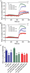
Figure 5. Effect of SOCE inhibitors ML9 and 2-APB on TG-sensitive SOCE in transfected cortical neurons.
(A–B) Cortical neurons were co-transfected with YFP-STIM1 and ORAI1 or YFP-STIM2 and ORAI1 or nontransfected (NT). Experiments were performed as described in Figure 4, but (A) 100 µM ML9 or (B) 50 µM 2-APB was added before store depletion with TG in the presence of inhibitors. Ca2+ (2 mM) was then added to assess SOCE. Each trace represents the average response of cells measured on a single coverslip from three independent experiments. F340/F380 values beginning just before the addition of TG were normalized to the same values (1). (C) The average increase in Ca2+ entry observed after the addition of 2 mM CaCl2 to transfected or nontransfected cells in the absence or presence of the SOCE inhibitors from experiments performed as described in (A–B). The data show the peak of the F340/F380 ratio after extracellular Ca2+ was added. ***p<0.001; ns, not significant (ANOVA). Error bars represent the standard error from three independent experiments.
Figure 6
Figure 6. Analysis of spontaneous store repletion in transfected neurons.
(A) Cytosolic Ca2+ measurements were performed in cells overexpressing YFP-STIM1 ± ORAI1 or YFP-STIM2 ± ORAI1 or in nontransfected (NT) cells. The experiments started in the presence of 2 mM CaCl2, followed by transfer to Ca2+-free medium. A buffer was then supplemented with 2 mM CaCl2 for 5 min to monitor intracellular Ca2+ restoration. Finally, 50 µM 2-APB was added. Raw (not normalized) traces are shown, which represent the average of at least 30–50 cells from two independent experiments. (B) Summary of basal Ca2+ at the beginning of the experiment. Data are shown as the Delta Ratio values, which were calculated as the difference between the F340/F380 ratio after extracellular Ca2+ was removed and its maximum level at the beginning of the experiment. (C–D) Analysis of constitutive calcium entry in transfected cortical neurons. Traces were normalized to 1 and are shown as a Supplementary Material (Figure S2A). (C) The average increases in Ca2+ entry (peak Ca2+ rise) observed after the addition of 2 mM CaCl2 to transfected cells compared with nontransfected cells are shown. (D) Statistical evaluation of the integrated Ca2+ responses (area under the curve [AUC]) is shown. (E) Effect of 2-APB on Ca2+ responses (peak Ca2+ rise). Traces normalized to 1 are shown as Supplementary Material (Figure S2B). The average Ca2+ responses to 50 µM 2-APB in 2 mM Ca2+ in cells transfected or nontransfected are shown. (B–E) *p<0.05, ***p<0.001, compared with control; ns, not significant (ANOVA). Bars indicate mean ± SD from at least 30–50 cells measured in two independent experiments.

References

    1. Putney JW., Jr A model for receptor-regulated calcium entry. Cell Calcium. 1986;7:1–12. - PubMed
    1. Blaustein MP, Golovina VA. Structural complexity and functional diversity of endoplasmic reticulum Ca(2+) stores. Trends Neurosci. 2001;24:602–608. - PubMed
    1. Berridge MJ, Irvine RF. Inositol trisphosphate, a novel second messenger in cellular signal transduction. Nature. 1984;312:315–321. - PubMed
    1. Berridge MJ, Bootman MD, Roderick HL. Calcium signalling: dynamics, homeostasis and remodelling. Nat Rev Mol Cell Biol. 2003;4:517–529. - PubMed
    1. Liou J, Kim ML, Heo WD, Jones JT, Myers JW, et al. STIM is a Ca2+ sensor essential for Ca2+-store-depletion-triggered Ca2+ influx. Curr Biol. 2005;15:1235–1241. - PMC - PubMed

Publication types

MeSH terms

Substances

LinkOut - more resources