. 2011 Apr 26;6(4):e19310.
doi: 10.1371/journal.pone.0019310.

Differentiation generates paracrine cell pairs that maintain basaloid mouse mammary tumors: proof of concept

Affiliations

Differentiation generates paracrine cell pairs that maintain basaloid mouse mammary tumors: proof of concept

Soyoung Kim et al. PLoS One. .

Abstract

There is a paradox offered up by the cancer stem cell hypothesis. How are the mixed populations that are characteristic of heterogeneous solid tumors maintained at constant proportion, given their high, and different, mitotic indices? In this study, we evaluate a well-characterized mouse model of human basaloid tumors (induced by the oncogene Wnt1), which comprise mixed populations of mammary epithelial cells resembling their normal basal and luminal counterparts. We show that these cell types are substantially inter-dependent, since the MMTV LTR drives expression of Wnt1 ligand in luminal cells, whereas the functional Wnt1-responsive receptor (Lrp5) is expressed by basal cells, and both molecules are necessary for tumor growth. There is a robust tumor initiating activity (tumor stem cell) in the basal cell population, which is associated with the ability to differentiate into luminal and basal cells, to regenerate the oncogenic paracrine signaling cell pair. However, we found an additional tumor stem cell activity in the luminal cell population. Knowing that tumors depend upon Wnt1-Lrp5, we hypothesized that this stem cell must express Lrp5, and found that indeed, all the stem cell activity could be retrieved from the Lrp5-positive cell population. Interestingly, this reflects post-transcriptional acquisition of Lrp5 protein expression in luminal cells. Furthermore, this plasticity of molecular expression is reflected in plasticity of cell fate determination. Thus, in vitro, Wnt1-expressing luminal cells retro-differentiate to basal cell types, and in vivo, tumors initiated with pure luminal cells reconstitute a robust basal cell subpopulation that is indistinguishable from the populations initiated by pure basal cells. We propose this is an important proof of concept, demonstrating that bipotential tumor stem cells are essential in tumors where oncogenic ligand-receptor pairs are separated into different cell types, and suggesting that Wnt-induced molecular and fate plasticity can close paracrine loops that are usually separated into distinct cell types.


Conflict of interest statement

Competing Interests: The authors have declared that no competing interests exist.

Figures

Figure 1
Figure 1. Despite lineage-specific expression of Wnt1 by purified basal and luminal cell subpopulations, both cell types express Axin2, and both serve as tumor initiating cells.
(A) A representative MMTV-Wnt-1-induced tumor paraffin-embedded section stained with lineage-specific markers, keratin-8 (K8; luminal; red) and keratin-5 (K5; basal; blue), together with Ki67, a marker of cycling cells (green). Scale bar = 50 µm. (B) Flow cytometric separation of luminal and basal cells from an MMTV-Wnt1 induced tumor, based on their expression of EpCAM and CD49f (results are similar for at least 3 primary tumors; a representative gating tree is shown in Fig. S1). The purity of these fractions is shown in Fig. S3. (C, D) mRNA extracted from purified cell fractions was analyzed for relative expression of Wnt1 and the Wnt reporter, Axin2, by quantitative RT-PCR analysis (n = 2, triplicates). MG, mammary gland; Hyper, Wnt1-induced hyperplastic glands; BAS, basal cells; LUM, luminal cells; Lin−, CD45−, CD31− (lineage-negative) live cells (total un-separated cells, run through the cell sorter).
Figure 2
Figure 2. Lrp5 expression (acquired post-transcriptionally) correlates with the appearance of Axin2 in luminal cells.
(A) mRNA extracted from purified cell fractions was analyzed for relative expression of Lrp5 by quantitative RT-PCR analysis as described for Fig. 1. (Lrp6 mRNA expression is broadly similar to Lrp5, shown in Fig. S4) (B) Tumor cells were incubated with anti-Lrp5 antibody, and analyzed by flow cytometry, using an isotype-matched negative control to define Lrp5+ cells. These samples were also stained with EpCAM and CD49f antibodies, and the cell surface phenotypes combined to show the luminal (L) or basal (B) cell identity of Lrp5+ and Lrp5− cells. (C) Another example of this assay is shown in histogram form, to reveal the overall level of expression for Lrp5 in luminal and basal cells in normal and tumor cells. (D) Paraffin sections from normal and Wnt-induced mammary glands were stained to illustrate the relative rates of division (green, Ki67) of basal (blue, K5) and luminal (red, K8) cells. Note that the green color stain in the lumens is a common artifact associated with non-specific binding to sticky luminal proteins. Scale bar = 50 µm. (E) Axin2lacZ [MMTV-Wnt1] (and control Wnt1) glands were stained in whole mounts for the lacZ reporter, followed by embedding, sectioning and immunocytochemical assay of basal (K5+) and luminal (K8+) cells. The panel on the LHS shows there was no background when the Axin2lacZ reporter was not present. The samples were incubated in x-gal substrate (B, basal; L, luminal; as indicated). The pattern of staining of AxinlacZ was heterogeneous, in some areas, stain was basal-specific, in others, there was also light staining throughout the luminal population, in others, focalized clusters of luminal cells showed high expression. (F) Lrp5+ cells were assayed for their expression of CD61, because CD61+ cells have been shown to be enriched for TIC activity in this tumor model (Vaillant et al 2008). These markers showed high co-expression.
Figure 3
Figure 3. Functional evaluation of Wnt1-induced basal and luminal populations in vivo.
Tumors regenerated from limiting dilutions of different cell fractions (Lin− total population, luminal or basal cells) were compared with samples of parental tumors immunostained for constituent cell types with K5 (blue), K8 (red), and Ki67 (green). Scale bar = 50 µm. These tumors were also analyzed by flow cytometry (using EpCAM and CD49f expression) to confirm the relative fate allocation of tumor subpopulations.
Figure 4
Figure 4. Functional evaluation of Wnt1-induced luminal cells in culture.
To evaluate the differentiation potential of luminal cells from Wnt1-induced mammary glands, flow sorted luminal cells were placed into culture, and stained 4 days later for markers of cell fate (K5, SMA and K8), together with a nuclear counter-stain. Under these conditions, purified luminal cells (<1% K5-positive) from normal glands continue to express K8, and were 98.9% K5-negative after 4 days. However, purified luminal cells from MMTV-Wnt1 hyperplastic glands (also <1% K5-positive) show mixed fates after transfer to tissue culture, including 24% K5-positive cells. Similarly, cultures of purified luminal cells from MMTV-Wnt1-induced tumors develop a similar proportion (21%) of K5-positive cells (n = 2, triplicates). Note that some cells are K5-positive, some are SMA-positive and most are positive for both. For BALB/c MECs, this is typical of the basal/myoepithelial lineage (in vivo and directly after isolation), and probably reflects an evolution of phenotype during differentiation (cells expressing SMA alone are most differentiated; data not shown). The corresponding cultures of basal cells from all three types of MEC population are shown in Fig. S7. Scale bar = 50 µm.
Figure 5
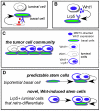
Figure 5. Summary diagram.
(A) In normal mammary epithelium, the Wnt source that maintains Lrp5+ mammary stem cells is indicated by a red-colored nucleus in a non-epithelial cell type, inferred from . (B) For Wnt1-induced mammary tumors, we have illustrated a paracrine signaling network that depends upon interactive basal and luminal cells. Wnt1 ligand and Lrp5 receptor are necessary and sufficient to maintain tumors (Lindvall et al., 2006). Wnt1, under the control of the MMTV-LTR sequence, is expressed by luminal cells (this cis sequence is only upregulated in luminal cells). Lrp5 protein is typically expressed by basal cells. (C) Cells that comprise the tumor cell community include (Lrp5+) basal cells that can differentiate to luminal cells to provide a ligand-expressing luminal partner. We have shown that some of these luminal cells also express Lrp5. (D) All cells with stem cell activity express Lrp5 and are able to regenerate robust mixtures of basal and luminal cells that support tumor growth by paracrine interactions. Thus, basal cells are predictable stem cells, since they differentiate to Wnt1-expressing luminal cells, whereas luminal cells are novel, Wnt-induced stem cells that express sufficient Lrp5 to enable their survival, but also show fate plasticity, to enable retro-differentiation to basal cells.

Similar articles

Cited by

References

    1. Wicha MS, Liu S, Dontu G. Cancer stem cells: an old idea–a paradigm shift. Cancer Res. 2006;66:1883–1890; discussion 1895–1886. - PubMed
    1. Carey LA, Dees EC, Sawyer L, Gatti L, Moore DT, et al. The triple negative paradox: primary tumor chemosensitivity of breast cancer subtypes. Clin Cancer Res. 2007;13:2329–2334. - PubMed
    1. Badve S, Dabbs DJ, Schnitt SJ, Baehner FL, Decker T, et al. Basal-like and triple-negative breast cancers: a critical review with an emphasis on the implications for pathologists and oncologists. Mod Pathol 2010 - PubMed
    1. Rakha EA, Elsheikh SE, Aleskandarany MA, Habashi HO, Green AR, et al. Triple-negative breast cancer: distinguishing between basal and nonbasal subtypes. Clin Cancer Res. 2009;15:2302–2310. - PubMed
    1. Cheang MC, Voduc D, Bajdik C, Leung S, McKinney S, et al. Basal-like breast cancer defined by five biomarkers has superior prognostic value than triple-negative phenotype. Clin Cancer Res. 2008;14:1368–1376. - PubMed

Publication types

MeSH terms

LinkOut - more resources