. 2011 Apr 20:5:26.
doi: 10.3389/fnana.2011.00026. eCollection 2011.

Somatotopic organization of the primate Basal Ganglia

Affiliations

Somatotopic organization of the primate Basal Ganglia

Atsushi Nambu. Front Neuroanat. .

Abstract

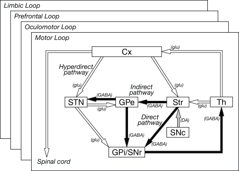
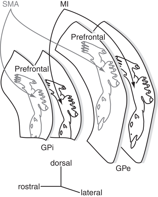
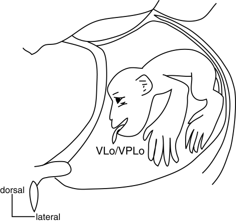
Somatotopic organization is a fundamental and key concept to understand how the cortico-basal ganglia loop works. It is also indispensable knowledge to perform stereotaxic surgery for movement disorders. Here I would like to describe the somatotopic organization of the basal ganglia, which consist of the striatum, subthalamic nucleus, globus pallidus, and substantia nigra. Projections from motor cortical regions representing different body parts terminate in different regions of these nuclei. Basal ganglia neurons respond not only to the stimulation of the corresponding regions of the motor cortices, but also to active and passive movements of the corresponding body parts. On the basis of these anatomical and physiological findings, somatotopic organization can be identified in the motor territories of these nuclei in the basal ganglia. In addition, projections from functionally interrelated cortical areas partially converge through the cortico-basal ganglia loop, but nevertheless the somatotopy is still preserved. Disorganized somatotopy may explain, at least in part, the pathophysiology of movement disorders, such as Parkinson's disease and dystonia.

Keywords: globus pallidus; movement disorders; somatotopy; striatum; substantia nigra; subthalamic nucleus.


Figures

Figure 1
Figure 1
Basic circuitry of the basal ganglia. Open and filled arrows indicate excitatory and inhibitory projections, respectively. Cx, cerebral cortex: DA, dopamine, GABA, gamma-aminobutyric acid; glu, glutamate; GPe and GPi, external and internal segments of the globus pallidus; SNc, substantia nigra pars compacta; SNr substantia nigra pars reticulata; STN, subthalamic nucleus; Str, striatum; Th, thalamus. Modified from Nambu et al. (2002b).
Figure 2
Figure 2
Information processing in the basal ganglia. (A) Parallel processing hypothesis. Information originating from different areas (a, b, c) of the cerebral cortex is processed independently in the different parts of the basal ganglia, and returns to the original cortical areas. (B) Information convergence hypothesis. Information originating from different cortical areas converges and is integrated in the basal ganglia, and integrated information returns to all the cortical areas. (C) Intermediate hypothesis between parallel processing and information convergence hypotheses, which is supported by recent studies.
Figure 3
Figure 3
Somatotopy of the putamen. (A) Somatotopy of the putamen is schematically shown in a frontal section. In the caudal aspect of the putamen, the lateral part receives somatotopic inputs from the primary motor cortex (MI), and the medial part from the supplementary motor area (SMA). The somatotopy in the SMA territory is located dorsomedially to that in the MI territory. Projections from the orofacial, forelimb and hindlimb regions of the MI and SMA converge in the medio-lateral central zone in the putamen. The most dorsomedial part receives inputs from the prefrontal cortex. Modified from Nambu et al. (2002a) (B) Input from motor cortices to the putamen is schematically shown in a horizontal section. CMAc and CMAr, caudal and rostral parts of the cingulate motor area; PMdc, PMdr, and PMvc, caudal part of dorsal premotor cortex, rostral part of dorsal premotor cortex, and caudal part of ventral premotor cortex. Modified from Takada et al. (2001).
Figure 4
Figure 4
Somatotopy of the subthalamic nucleus (STN) and substantia nigra (SN). (A) Somatotopy of the STN is shown in a frontal section. In the dorsal part of the STN, the lateral part receives somatotopic inputs from the MI, and the medial part from the SMA. The MI also projects partly to the medial part, and the SMA to the lateral part. Ventral to the motor territory, there exist the oculomotor and prefrontal territories. The most medial part is occupied by the limbic territory. Modified from Nambu et al. (2002b). (B) Input from motor cortices to the STN is schematically shown in a horizontal section. Modified from Takada et al. (2001). (C) Somatotopy of the SNr is shown in a frontal section. The orofacial region of the SNr is a continuation of that of the GPi (see Figure 5). Ventral to the motor territory, there exist the oculomotor and prefrontal territories.
Figure 5
Figure 5
Somatotopy of the external and internal segments of the globus pallidus (GPe and GPi). The GPe and GPi have their own somatotopic representations. The somatotopy in the SMA territory is located rostrodorsally to that in the MI territory. Two territories overlap partly in the rostro-caudal zone. Dorsal one-third of the GPe/GPi is occupied by the prefrontal territory.
Figure 6
Figure 6
Somatotopy of the thalamus. Somatotopy of the thalamus is shown in a frontal section. Both the oral part of the ventrolateral nucleus of the thalamus (VLo) receiving pallidal projections and the oral part of the ventroposterolateral nucleus of the thalamus (VPLo) receiving cerebellar projections have their own somatotopic representations. Their somatotopic representations are continuous rostrocaudally. Modified from Asanuma et al. (1983).

References

    1. Akkal D., Dum R. P., Strick P. L. (2007). Supplementary motor area and presupplementary motor area: targets of basal ganglia and cerebellar output. J. Neurosci. 27, 10659–1067310.1523/JNEUROSCI.3134-07.2007 - DOI - PMC - PubMed
    1. Alexander G. E., Crutcher M. D. (1990a). Functional architecture of basal ganglia circuits: neural substrates of parallel processing. Trends Neurosci. 13, 266–27110.1016/0166-2236(90)90107-L - DOI - PubMed
    1. Alexander G. E., Crutcher M. D. (1990b). Preparation for movement: neural representations of intended direction in three motor areas of the monkey. J. Neurophysiol. 64, 133–150 - PubMed
    1. Alexander G. E., DeLong M. R. (1985). Microstimulation of the primate neostriatum. II. Somatotopic organization of striatal microexcitable zones and their relation to neuronal response properties. J. Neurophysiol. 53, 1417–1430 - PubMed
    1. Alexander G. E., DeLong M. R., Strick P. L. (1986). Parallel organization of functionally segregated circuits linking basal ganglia and cortex. Annu. Rev. Neurosci. 9, 357–381 - PubMed

LinkOut - more resources