. 2011 Apr 26;6(4):e18441.
doi: 10.1371/journal.pone.0018441.

Virulence evolution of the human pathogen Neisseria meningitidis by recombination in the core and accessory genome

Affiliations

Virulence evolution of the human pathogen Neisseria meningitidis by recombination in the core and accessory genome

Biju Joseph et al. PLoS One. .

Abstract

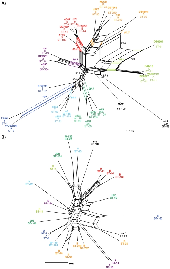
Background: Neisseria meningitidis is a naturally transformable, facultative pathogen colonizing the human nasopharynx. Here, we analyze on a genome-wide level the impact of recombination on gene-complement diversity and virulence evolution in N. meningitidis. We combined comparative genome hybridization using microarrays (mCGH) and multilocus sequence typing (MLST) of 29 meningococcal isolates with computational comparison of a subset of seven meningococcal genome sequences.

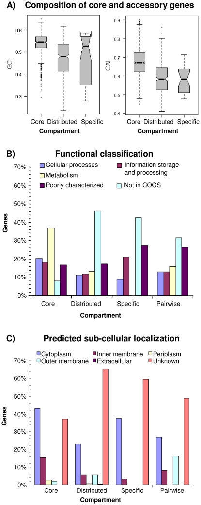
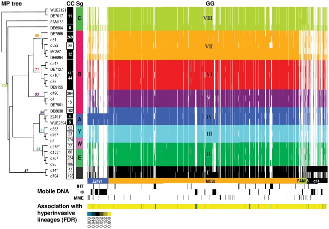
Principal findings: We found that lateral gene transfer of minimal mobile elements as well as prophages are major forces shaping meningococcal population structure. Extensive gene content comparison revealed novel associations of virulence with genetic elements besides the recently discovered meningococcal disease associated (MDA) island. In particular, we identified an association of virulence with a recently described canonical genomic island termed IHT-E and a differential distribution of genes encoding RTX toxin- and two-partner secretion systems among hyperinvasive and non-hyperinvasive lineages. By computationally screening also the core genome for signs of recombination, we provided evidence that about 40% of the meningococcal core genes are affected by recombination primarily within metabolic genes as well as genes involved in DNA replication and repair. By comparison with the results of previous mCGH studies, our data indicated that genetic structuring as revealed by mCGH is stable over time and highly similar for isolates from different geographic origins.

Conclusions: Recombination comprising lateral transfer of entire genes as well as homologous intragenic recombination has a profound impact on meningococcal population structure and genome composition. Our data support the hypothesis that meningococcal virulence is polygenic in nature and that differences in metabolism might contribute to virulence.


Conflict of interest statement

Competing Interests: The authors have declared that no competing interests exist.

Figures

Figure 1
Figure 1. Characterization of the meningococcal gene complement.
A) Boxplots comparing the GC content and CAI between core, distributed and strain-specific genes. Core genes have a significantly higher GC content than the distributed genes and also a significantly higher CAI than genes from the accessory genome comprising distributed and strain-specific genes (p<0.001, Wilcoxon test). B) Distribution of genes from the different genomic compartments also including pairwise differences between strains from the same CC among the major COG functional classes. There are significant differences in the functional composition between all four genomic compartments (p<0.01, χ2 test). Compared to the other genomic compartments the core genome is enriched for metabolic genes as well as genes involved in cellular processes, information storage and processing, respectively (p<0.01, Fisher's exact test). C) Histogram of the predicted subcellular localization of genes from the different genomic compartments. Again, there are significant differences between the different genomic compartments (p<0.01, χ2 test). In line with their housekeeping functions, core genes are localized primarily within the cytoplasm, the inner membrane and the periplasmic space whereas the distributed genes and genes that differ between strains from the same CC are in turn enriched for genes coding for outer membrane proteins (p<0.05, Fisher's exact test).
Figure 2
Figure 2. Clustering of strains based on the accessory genome.
A maximum parsimony (MP) tree is shown with bootstrap values at nodes used for grouping of strains into eight GGs from their gene content comprising 470 parsimony informative genes as revealed by mCGH. Strains with an asterisk next to their name have further been used for the in silico screening for intragenomic recombination in 1092 genes from the core genome as estimated via mCGH of the entire sample population. Next to the MP tree, the CCs and serogroups (Sg) of the respective strains are given with hypervirulent CCs in black boxes, and right to the Sgs a virtual array image displaying the presence and absence of 1679 genes is shown. Strains from the same serogroup have in general highly similar gene content, and strains from the same CC always belong to the same GG. In turn, a GG can comprise strains from different CCs, and with the exception of the two serogroup W-135 strains split between GG-II and GG-III and the serogroup B strain DE8638, GGs always comprise strains from the same serogroup. However, no two strains have exactly the same gene content (see also Figure 1 for the functional profile of genes differently present among otherwise identical pairs of strains). Right below the virtual array, the spotted genes are color coded according to the source genome (representing the genomes of strain Z2491, MC58, FAM18 and α14), and the presence of putatively mobile DNA is depicted below with IHT-B, IHT-C, IHT-E as well as the λ prophage denoted as B, C, E and λ, respectively, in the respective lanes (Abbreviations: IHT, island of horizontal transfer; Φ: prophage; MME, minimal mobile element). At the lower margin, the FDR for the association with hyperinvasive lineages is color coded for each gene with genes having a FDR<0.05 depicted in blue.
Figure 3
Figure 3. Comparison of strain clustering based on mCGH and MLST.
A) Neighbor-net representation of gene distances based on the distribution of genes from the accessory genome. Strain names as well as the defining splits having a bootstrap support of greater than 85% are printed in the same colors as in Figure 2. Clustering based on gene content distance and maximum parsimony results in the same eight genome groups. Serogroup B and C strains as well as the two cnl strains are separated from the other four serogroups based on gene content, and the cycles within the serogroup B/C subgraph indicate a high rate of gene transfer among these strains. Strains from the same CC always reside on common splits. B) Neighbor-net representation based on the concatenated alignments of the seven housekeeping gene fragments form the meningococcal core genome used for MLST comprising 383 parsimony informative sites. A MLSA-based clustering of strains results in the separation of strains from the same serogroup or belonging to the same GGs. The large number of cycles in the graph indicates a high rate of homologous recombination among the genes used for MLST and that a MLSA-based reconstruction of the deep phylogeny of these strains is not possible.

Similar articles

Cited by

References

    1. Claus H, Maiden MC, Wilson DJ, McCarthy ND, Jolley KA, et al. Genetic analysis of meningococci carried by children and young adults. J Infect Dis. 2005;191:1263–1271. - PubMed
    1. Yazdankhah SP, Kriz P, Tzanakaki G, Kremastinou J, Kalmusova J, et al. Distribution of serogroups and genotypes among disease-associated and carried isolates of Neisseria meningitidis from the Czech Republic, Greece, and Norway. J Clin Microbiol. 2004;42:5146–5153. - PMC - PubMed
    1. Stephens DS, Greenwood B, Brandtzaeg P. Epidemic meningitis, meningococcaemia, and Neisseria meningitidis. Lancet. 2007;369:2196–2210. - PubMed
    1. Frosch M, Vogel U. Structure and genetics of the meningococcal capsule. In: Frosch M, Maiden MC, editors. Handbook of Meningococcal Disease. Weinheim, Germany: Wiley-VCH; 2006. pp. 145–162.
    1. Harrison LH, Trotter CL, Ramsay ME. Global epidemiology of meningococcal disease. Vaccine. 2009;27(Suppl 2):B51–63. - PubMed

Publication types