. 2011 Apr 26;6(4):e18995.
doi: 10.1371/journal.pone.0018995.

A recombinant influenza A virus expressing domain III of West Nile virus induces protective immune responses against influenza and West Nile virus

Affiliations

A recombinant influenza A virus expressing domain III of West Nile virus induces protective immune responses against influenza and West Nile virus

Byron E E Martina et al. PLoS One. .

Abstract

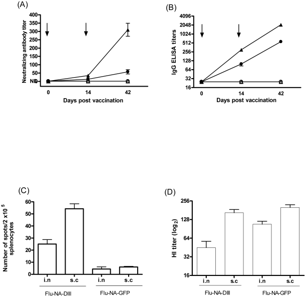
West Nile virus (WNV) continues to circulate in the USA and forms a threat to the rest of the Western hemisphere. Since methods for the treatment of WNV infections are not available, there is a need for the development of safe and effective vaccines. Here, we describe the construction of a recombinant influenza virus expressing domain III of the WNV glycoprotein E (Flu-NA-DIII) and its evaluation as a WNV vaccine candidate in a mouse model. FLU-NA-DIII-vaccinated mice were protected from severe body weight loss and mortality caused by WNV infection, whereas control mice succumbed to the infection. In addition, it was shown that one subcutaneous immunization with 10(5) TCID(50) Flu-NA-DIII provided 100% protection against challenge. Adoptive transfer experiments demonstrated that protection was mediated by antibodies and CD4+T cells. Furthermore, mice vaccinated with FLU-NA-DIII developed protective influenza virus-specific antibody titers. It was concluded that this vector system might be an attractive platform for the development of bivalent WNV-influenza vaccines.


Conflict of interest statement

Competing Interests: None of the authors declare conflict of interest apart from Albert Osterhaus, who is a part-time employee (CEO) of Viroclinics B.V. (for details go to www.erasmusmc.nl) and Gunther Spohn who is an employee of Cytos Biotechnology. Guus Rimmelzwaan has an affiliation with Viroclinics BV as a consultant. The stated competing interests do not alter the authors' adherence to all the PLoS ONE policies on sharing data and materials.

Figures

Figure 1
Figure 1. Characterization of recombinant influenza A viruses.
RT-PCR analysis of Flu-NA-GFP and Flu-NA-DIII RNA extracted from MDCK cells 20 hours after infection (A). Amplicons were separated in 1% agarose gel. For amplification WNV-DIII (lanes 2 and 3) and influenza A (lanes 6 and 7) virus-specific primers were used. Expression of DIII was analyzed by Western blot analysis (B). Viral proteins in lysates of infected cells or in sucrose gradient purified virus preparations were separated by SDS-PAGE and transferred to PDVF membranes, which were incubated with a DIII specific monoclonal antibody 7H2 (upper panel) or an influenza virus NP specific monoclonal antibody (ATCC, clone HB65; lower panel). Expression of DIII was also confirmed by immuno-staining of MDCK cell infected with FLU-NA-GFP and FLU-NA-DIII (moi = 0.01) with NP- and DIII-specific antibodies as indicated (C).
Figure 2
Figure 2. Induction of WNV-specific immune responses by vaccination with FLU-NA-DIII.
WNV-neutralizing antibodies were detected by virus neutralization assay (A) and DIII-specific IgG antibodies by ELISA (B) in serum obtained from mice vaccinated with FLU-NA-DIII i.n. (•) or s.c. (▴) and FLU-NA-GFP i.n. (○) or s.c. (▵) at the indicated time points. Arrows indicate time points of vaccination. The data are expressed as average titers per group (n = 10) ± SD. Cellular DIII-specific responses were determined by IFN- γ ELISPOT assay (C). Splenocytes obtained on day 42 were stimulated with 10 µM peptide (VNPFVSVATANAKVL) and the numbers of cells producing IFN-γ per 2×10⁵ cells were determined in mice vaccinated with FLU-NA-DIII or FLU-NA-GFP as indicated. Each experiment was performed twice in triplicate. Results are indicated as mean ± standard deviation. Induction of Hemagglutination titers after immunization with FLU-NA-DIII and FLU-NA-GFP. Mice (n = 8) were immunized intranasally (i.n.) or subcutaneously (s.c.) with Flu-NA-DIII or FLU-NA-GFP (D).
Figure 3
Figure 3. Loss of body weight after challenge-infection with WNV.
Mice (n = 8) were vaccinated intranasally with Flu-NA-DIII (•) or Flu-NA-GFP (○) or by the subcutaneous route (▴ and ▵, respectively). The daily weights of each animal were calculated compared to their respective weight on the day of challenge, and data are shown as the average percentage of initial weight for each group. Error bars represent the standard error for all samples available at that time point. Subsequently, the mice were challenged subcutaneously with 106 TCID50 WNV-NY99 and weighed daily. The mean body weight is expressed as the percentage of the body weight before challenge infection (A). The survival rates of mice after challenge infection with WNV-NY99 are depicted as Kaplan-Meier survival curves (B). The difference in survival rate between Flu-NA-DIII and Flu-NA-GFP vaccinated mice was statistically significant as determined by the logrank test. The symbols for the respective groups are the same as in panel A.
Figure 4
Figure 4. Protection against challenge infection is mediated by humoral and CD4+T cell responses.
Recipient mice received serum (A and B) CD4+T cells (C and D) or CD8+T cells (E and F) obtained from mice that were vaccinated with FLU-NA-DIII (closed symbols) or FLU-NA-GFP (open symbols) by the i.n route (• and ○ respectively) or s.c. (▴ and ▵ respectively) and were subsequently infected with 100 TCID50 WNV-NY99. Loss of body weight (A,C and E) and virus titers in the brain were determined eight days post challenge infection (B, D and F). The results represent the mean values of groups of five mice. Error bars indicate the standard deviation; * indicates a statistically significant difference compared to control groups receiving serum or T cells from FLU-NA-GFP vaccinated mice (determined by t test). The daily weights of each animal were calculated compared to their respective weight on the day of challenge, and data are shown as the average percentage of initial weight for each group (A, C, E). Error bars represent the standard error for all samples available at that time point.

Similar articles

Cited by

References

    1. Mostashari F, Bunning ML, Kitsutani PT, Singer DA, Nash D, et al. Epidemic West Nile encephalitis, New York, 1999: results of a household-based seroepidemiological survey. Lancet. 2001;358:261–264. - PubMed
    1. Pawelec G, Rehbein A, Haehnel K, Merl A, Adibzadeh M. Human T-cell clones in long-term culture as a model of immunosenescence. Immunol Rev. 1997;160:31–42. - PubMed
    1. Ligthart GJ, Corberand JX, Fournier C, Galanaud P, Hijmans W, et al. Admission criteria for immunogerontological studies in man: the SENIEUR protocol. Mech Ageing Dev. 1984;28:47–55. - PubMed
    1. Fleming DM, Elliot AJ. Lessons from 40 years' surveillance of influenza in England and Wales. Epidemiol Infect. 2008;136:866–875. - PMC - PubMed
    1. Gubler DJ. The continuing spread of West Nile virus in the western hemisphere. Clin Infect Dis. 2007;45:1039–1046. - PubMed

Publication types

MeSH terms