. 2011 Apr 25;6(4):e18989.
doi: 10.1371/journal.pone.0018989.

Full-length L1CAM and not its Δ2Δ27 splice variant promotes metastasis through induction of gelatinase expression

Affiliations

Full-length L1CAM and not its Δ2Δ27 splice variant promotes metastasis through induction of gelatinase expression

Stephanie Hauser et al. PLoS One. .

Abstract

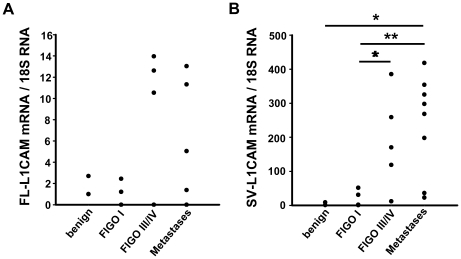
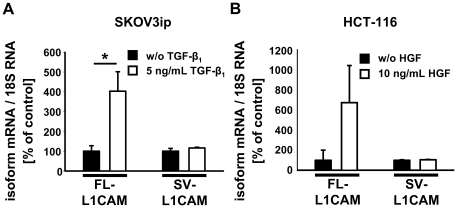
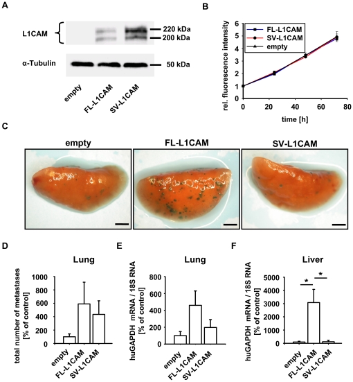
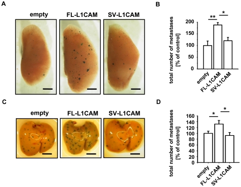
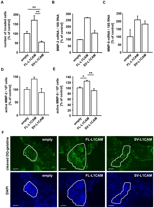
Tumour-specific splicing is known to contribute to cancer progression. In the case of the L1 cell adhesion molecule (L1CAM), which is expressed in many human tumours and often linked to bad prognosis, alternative splicing results in a full-length form (FL-L1CAM) and a splice variant lacking exons 2 and 27 (SV-L1CAM). It has not been elucidated so far whether SV-L1CAM, classically considered as tumour-associated, or whether FL-L1CAM is the metastasis-promoting isoform. Here, we show that both variants were expressed in human ovarian carcinoma and that exposure of tumour cells to pro-metastatic factors led to an exclusive increase of FL-L1CAM expression. Selective overexpression of one isoform in different tumour cells revealed that only FL-L1CAM promoted experimental lung and/or liver metastasis in mice. In addition, metastasis formation upon up-regulation of FL-L1CAM correlated with increased invasive potential and elevated Matrix metalloproteinase (MMP)-2 and -9 expression and activity in vitro as well as enhanced gelatinolytic activity in vivo. In conclusion, we identified FL-L1CAM as the metastasis-promoting isoform, thereby exemplifying that high expression of a so-called tumour-associated variant, here SV-L1CAM, is not per se equivalent to a decisive role of this isoform in tumour progression.


Conflict of interest statement

Competing Interests: The authors have read the journal's policy and have the following conflicts: Ulrich Weidle is employed by Roche Diagnostics GmbH. There are no patents, products in development or marketed products to declare. This does not alter the authors' adherence to all the PLoS ONE policies on sharing data and materials.

Figures

Figure 1
Figure 1. Both FL-L1CAM and SV-L1CAM were expressed in benign and malignant tumours of human ovarian carcinoma patients.
Expression of FL-L1CAM (A) and SV-L1CAM mRNA (B) was analyzed in benign and malignant ovarian tumours and their peritoneal metastases. Mean of relative target gene mRNA vs. 18S rRNA ± SEM: A. Values obtained for FL-L1CAM; benign tumours: 1.85±0.85, n = 2; FIGO I: 0.73±0.49, n = 5; FIGO III or FIGO IV: 5.30±2.53, n = 7; metastases: 2.57±1.37, n = 12 (p = 0.664, as determined by Kruskal-Wallis One Way ANOVA on Ranks). B. Values obtained for SV-L1CAM; benign tumours: 4.99±3.99, n = 2; FIGO I: 17.33±10.41, n = 5; FIGO III or FIGO IV: 189.13±63.25, n = 5; metastases: 240.21±51.18, n = 8 (all groups: *p = 0.015, as determined by Kruskal-Wallis One Way ANOVA on Ranks and subsequent post hoc comparison using Holm-Sidak method; pairwise comparisons (unadjusted p values): FIGO I vs. benign: p = 0.903; FIGO III or FIGO IV vs. benign: p = 0.084; metastases vs. benign: *p = 0.024; FIGO III/IV vs. FIGO I: *p = 0.037; metastases vs. FIGO I: **p = 0.005; metastases vs. FIGO III/IV: p = 0.465).
Figure 2
Figure 2. Expression of L1CAM splice variants was deregulated in carcinoma cells upon exposure to pro-metastatic factors.
Mean FL-L1CAM or SV-L1CAM mRNA levels ± SEM (columns ± bars) after incubation with pro-metastatic factors. Target gene mRNA levels were normalized to 18S rRNA levels and the means of the relative reference group without incubation with a pro-metastatic factor were set as 100%. A. 1×10⁵ SKOV3ip-lacZ ovarian carcinoma cells were incubated for 48 h with or without 5 ng/ml of recombinant TGF-β1 (recTGF-β1). FL-L1CAM/Ø: 100.0%±27.6%, n = 9 cell pools; FL-L1CAM/recTGF-β1: 403.5%±98.1%, n = 9 cell pools; SV-L1CAM/Ø: 100.0%±14.2%, n = 9 cell pools; SV-L1CAM/recTGF-β1: 116.2%±4.0%, n = 9 cell pools (FL-L1CAM, SKOV3ip-lacZ+recTGF-β1 vs. SKOV3ip-lacZ−recTGF-β1: **p = 0.005; SV-L1CAM, SKOV3ip-lacZ+recTGF-β1 vs. SKOV3ip-lacZ−recTGF-β1: p = 0.266, as determined by Wilcoxon Signed Rank test). B. 1×10⁵ HCT-116 colon carcinoma cells were incubated for 10 days with or without 10 ng/ml of recombinant HGF (recHGF). FL-L1CAM/Ø: 100.0%±100.0%, n = 9 cell pools; FL-L1CAM/recHGF: 673.9%±369.7%, n = 9 cell pools; SV-L1CAM/Ø: 100.0%±4.6%, n = 9 cell pools; SV-L1CAM/recHGF: 104.9%±2.7%, n = 9 cell pools (FL-L1CAM, HCT-116+recHGF vs. HCT-116−recHGF: p = 0.313; SV-L1CAM, HCT-116+recHGF vs. HCT-116−recHGF: p = 0.250, as determined by Wilcoxon Signed Rank Test).
Figure 3
Figure 3. FL-L1CAM promoted the metastatic potential of HT1080lacZ-K15 human fibrosarcoma cells.
HT1080lacZ-K15 cells were transfected with retroviruses coding for FL-L1CAM cDNA or SV-L1CAM cDNA or with an empty vector without transgene. A. Western Blot analysis of L1CAM showed that HT1080lacZ-K15 cells did not express any L1CAM isoform endogenously, whereas expression of FL-L1CAM or SV-L1CAM was detected after gene transfer. The two bands at 220 kDa and 200 kDa are due to differential glycosylation status of L1CAM. B. alamarBlue® proliferation assay did not document any difference in proliferation between the different HT1080lacZ-K15 cell lines. Mean cell number ± SEM (dots ± bars). The mean of the 0 h value within each group was set as 1. Empty: 0 h: 1.000±0.046, 24 h: 2.039±0.037, 48 h: 3.457±0.203, 72 h: 4.950±0.401; FL-L1CAM: 0 h: 1.000±0.014, 24 h: 1.965±0.066, 48 h: 3.458±0.101, 72 h: 4.797±0.164; SV-L1CAM: 0 h: 1.000±0.016, 24 h: 2.096±0.112, 48 h: 3.328±0.031, 72 h: 4.918±0.241. C to E. 21 days after inoculation of 1×10⁶ of the different HT1080lacZ-K15 cells, CD1nu/nu mice were sacrificed, and their lungs and livers were removed. C. X-Gal staining (indigoblue foci) of removed lungs. Representative surface images are presented (bars: 2 mm). D. Mean number of macrometastases in lungs ± SEM (columns ± bars). The mean of the reference group (empty) was set as 100%. Empty: 100.0%±43.3%, n = 6 mice; FL-L1CAM: 554.6%±324.1%, n = 4 mice; SV-L1CAM: 434.0%±202.8%, n = 6 mice (FL-L1CAM vs. empty: p = 0.067; SV-L1CAM vs. empty: p = 0.180; SV-L1CAM vs. FL-L1CAM: p = 0.610, as determined by Mann-Whitney Rank Sum test). E. Mean human GAPDH mRNA levels ± SEM (columns ± bars) in lungs. GAPDH-mRNA levels were normalized to 18S rRNA levels and the mean of the reference group (empty) was set as 100%. Empty: 100.0%±48.4%, n = 6 mice; FL-L1CAM: 459.1%±171.2%, n = 4 mice; SV-L1CAM: 197.1%±92.9%, n = 6 mice (FL-L1CAM vs. empty: p = 0.067; SV-L1CAM vs. empty: p = 0.485; SV-L1CAM vs. FL-L1CAM: p = 0.257, as determined by Mann-Whitney Rank Sum test). F. Mean human GAPDH mRNA levels ± SEM (columns ± bars) in livers. GAPDH mRNA levels were normalized to 18S rRNA levels and the mean of the reference group (empty) was set as 100%. Empty: 100.0%±56.9%, n = 6 mice; FL-L1CAM: 3078.7%±999.7%, n = 4 mice; SV-L1CAM: 121.5%±109.2%, n = 6 mice (FL-L1CAM vs. empty: *p = 0.010; SV-L1CAM vs. empty: p = 1.000; SV-L1CAM vs. FL-L1CAM: *p = 0.019, as determined by Mann-Whitney Rank Sum Test).
Figure 4
Figure 4. FL-L1CAM also promoted the metastatic potential of SKOV3ip-lacZ human ovarian carcinoma and L-CI.5s murine T-lymphoma cells.
SKOV3ip-lacZ (A and B) as well as L-CI.5s (C and D) cells were stably transduced with retroviruses coding for cDNA of FL-L1CAM or SV-L1CAM or with a retroviral vector without cDNA (empty). A and B. 26 days after inoculation of 1.0×10⁵ of the different SKOV3ip-lacZ cells, CD1nu/nu mice were sacrificed and their lungs were removed. A. X-Gal staining (indigoblue foci) of removed lungs. Representative surface images are presented (bars: 2 mm). B. Mean number of macrometastases in lungs ± SEM (columns ± bars). The mean of the reference group (empty) was set as 100%; n = 4 mice each. Empty: 100.0%±19.1%; FL-L1CAM: 187.8%±10.3%; SV-L1CAM: 120.6%±14.2% (all groups: **p = 0.006, as determined by Kruskal-Wallis One Way ANOVA on Ranks and subsequent post hoc comparison using Dunn's method; pairwise comparisons (unadjusted p values): FL-L1CAM vs. empty: **p = 0.002; SV-L1CAM vs. empty: p = 0.355; SV-L1CAM vs. FL-L1CAM: *p = 0.011). C. and D. 7 days after inoculation of 5×10³ of the different L-CI.5s cells, DBA/2 mice were sacrificed and their livers were removed. C. X-Gal staining (indigoblue foci) of removed livers. Representative surface images are presented (bars: 2 mm). D. Mean number of macrometastases in livers ± SEM (columns ± bars). The mean of the reference group (empty) was set as 100%. Empty: 100.0%±7.5%, n = 10 mice; FL-L1CAM: 131.8%±12.4%, n = 4 mice; SV-L1CAM: 92.6%±10.3%, n = 5 mice (FL-L1CAM vs. empty: *p = 0.029; SV-L1CAM vs. empty: p = 0.200; SV-L1CAM vs. FL-L1CAM: *p = 0.029, as determined by Mann-Whitney Rank Sum test).
Figure 5
Figure 5. FL-L1CAM over-expression correlated with increased invasive capacity, gelatinase expression and activity in vitro, and gelatinolytic activity in vivo.
HT1080lacZ-K15 cells were stably transduced with retroviruses coding for cDNA of FL-L1CAM or SV-L1CAM or with a retroviral vector without cDNA (empty). A. Mean number of invaded cells per image section ± SEM (columns ± bars) for the different HT1080lacZ-K15 cells. The mean of the reference group (empty) was set as 100%; n = 4. Empty: 100.0%±10.6%; FL-L1CAM: 170.6%±23.5%; SV-L1CAM: 54.3%±5.4% (FL-L1CAM vs. empty: p = 0.057; SV-L1CAM vs. empty: **p = 0.009; SV-L1CAM vs. FL-L1CAM: **p = 0.003; as determined by Mann-Whitney Rank Sum test). B . and C. Mean MMP-2 (B) and MMP-9 (C) mRNA expression levels in the different HT1080lacZ-K15 cells ± SEM (columns ± bars) as quantified by qRT-PCR. The mean of the reference group (empty) was set as 100%; n = 3. B. Empty: 100.0%±2.7%; FL-L1CAM: 269.1%±3.0%; SV-L1CAM: 150.4%±37.6% (FL-L1CAM vs. empty: p = 0.100; SV-L1CAM vs. empty: p = 0.100; SV-L1CAM vs. FL-L1CAM: p = 0.100; as determined by Mann-Whitney Rank Sum Test). C. Empty: 100.0%±45.4%; FL-L1CAM: 207.7%±25.0%; SV-L1CAM: 180.7%±20.9% (FL-L1CAM vs. empty: p = 0.200; SV-L1CAM vs. empty: p = 0.200; SV-L1CAM vs. FL-L1CAM: p = 0.400; as determined by Mann-Whitney Rank Sum Test). Mean MMP-2 (D) and MMP-9 (E) activity of the different HT1080lacZ-K15 cells ± SEM (columns ± bars). The mean of the reference group (empty) was set as 100%. D. Empty: 100.0%±7.3%; FL-L1CAM: 140.4%±7.9%; SV-L1CAM: 87.5%±15.9%; n = 3 (FL-L1CAM vs. empty: p = 0.100; SV-L1CAM vs. empty: p = 0.700; SV-L1CAM vs. FL-L1CAM: p = 0.100; as determined by Mann-Whitney Rank Sum test). E. Empty: 100.0%±3.5%; FL-L1CAM: 124.5%±3.3%; SV-L1CAM: 90.0%±7.0%; n = 3 (all groups: *p = 0.007, as determined by Kruskal-Wallis One Way ANOVA on Ranks and subsequent post hoc comparison using Dunn's method; pairwise comparisons (unadjusted p values): FL-L1CAM vs. empty: *p = 0.013; SV-L1CAM vs. empty: p = 0.201; SV-L1CAM vs. FL-L1CAM: **p = 0.003). F. 21 days after inoculation of 1×10⁶ of the different HT1080lacZ-K15 cells, CD1nu/nu mice were sacrificed and their lungs were removed. In situ zymography was performed on cryo-sections of lungs bearing metastases of the different HT1080lacZ-K15 cells. Representative images are presented (bars: 50 µm; upper row (green signal): degraded DQ-gelatine; lower row (blue signal): DAPI counter-staining).
Figure 6
Figure 6. Differential endosomal sorting of FL-L1CAM compared to SV-L1CAM in HT1080lacZ-K15 human fibrosarcoma cells.
A. HT1080lacZ-K15 cells overexpressing FL-L1CAM before and after antibody-induced internalization. B. and C. HT1080lacZ-K15 cells 60 min chased at 37°C co-expressing FL-L1CAM or SV-L1CAM together with fluorophore-tagged LAMP1, KIF16B, or TGN38. Arrows indicate internalized L1CAM in selected vesicles that co-localized with compartment markers (Scale bars: 10 µm (A to C)). D. Relative distribution of L1CAM-labeled vesicles in lysosomes (GFP-LAMP1: SV-L1CAM: 24.0%±4.2% SEM; FL-L1CAM: 18.6±4.0% SEM; *p = 0.022, n = 30 each), early endosomes (YFP-KIF16B: SV-L1CAM: 43.3%±5.8% SEM; FL-L1CAM: 53.4%±5.1% SEM; p = 0.194, n = 30 each), retrograde and recycling endosomes (YFP-TGN38: SV-L1CAM: 35.5%±6.3% SEM; FL-L1CAM: 54.7%±5.2% SEM; **p = 0.002, n = 20 each) Data was calculated from three (FL- or SV-L1CAM+GFP-LAMP1, n = 30 each) or two independent experiments (FL- or SV-L1CAM+YFP-TGN38, n = 20 each) and displayed as means ± SEM (columns ± bars), p values were determined by Mann-Whitney Rank Sum Test.

Similar articles

Cited by

References

    1. Moos M, Tacke R, Scherer H, Teplow D, Fruh K, et al. Neural adhesion molecule L1 as a member of the immunoglobulin superfamily with binding domains similar to fibronectin. Nature. 1988;334:701–703. - PubMed
    1. Reid RA, Hemperly JJ. Variants of human L1 cell adhesion molecule arise through alternate splicing of RNA. J Mol Neurosci. 1992;3:127–135. - PubMed
    1. Breitbart RE, Andreadis A, Nadal-Ginard B. Alternative splicing: a ubiquitous mechanism for the generation of multiple protein isoforms from single genes. Annu Rev Biochem. 1987;56:467–495. - PubMed
    1. Ghigna C, Valacca C, Biamonti G. Alternative splicing and tumor progression. Curr Genomics. 2008;9:556–570. - PMC - PubMed
    1. Pajares MJ, Ezponda T, Catena R, Calvo A, Pio R, et al. Alternative splicing: an emerging topic in molecular and clinical oncology. Lancet Oncol. 2007;8:349–357. - PubMed

Publication types

MeSH terms

Substances