Comparative Study
. 2011 Oct 3;8(5):1573-81.
doi: 10.1021/mp2001704. Epub 2011 May 24.

Induced pluripotent stem (iPS) cells repair and regenerate infarcted myocardium

Affiliations
Comparative Study

Induced pluripotent stem (iPS) cells repair and regenerate infarcted myocardium

Dinender K Singla et al. Mol Pharm. .

Abstract

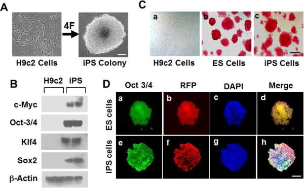
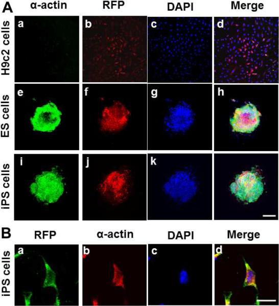
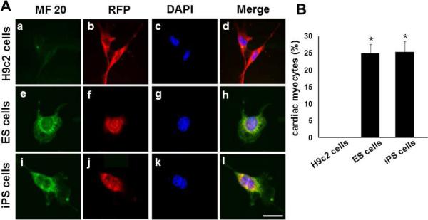
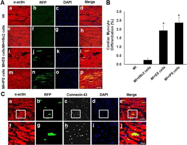
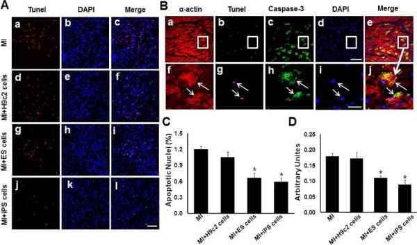
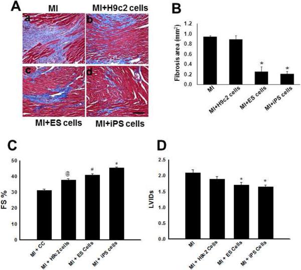
Cardiac myocyte differentiation reported thus far is from iPS cells generated from mouse and human fibroblasts. However, there is no article on the generation of iPS cells from cardiac ventricular specific cell types such as H9c2 cells. Therefore, whether transduced H9c2 cells, originally isolated from embryonic cardiac ventricular tissue, will be able to generate iPS cells and have the potential to repair and regenerate infarcted myocardium remains completely elusive. We transduced H9c2 cells with four stemness factors, Oct3/4, Sox2, Klf4, and c-Myc, and successfully reprogrammed them into iPS cells. These iPS cells were able to differentiate into beating cardiac myocytes and positively stained for cardiac specific sarcomeric α-actin and myosin heavy chain proteins. Following transplantation in the infarcted myocardium, there were newly differentiated cardiac myocytes and formation of gap junction proteins at 2 weeks post-myocardial infarction (MI), suggesting newly formed cardiac myocytes were integrated into the native myocardium. Furthermore, transplanted iPS cells significantly (p < 0.05) inhibited apoptosis and fibrosis and improved cardiac function compared with MI and MI+H9c2 cell groups. Moreover, our iPS cell derived cardiac myocyte differentiation in vitro and in vivo was comparable to embryonic stem cells in the present study. In conclusion we report for the first time that we have H9c2 cell-derived iPS cells which contain the potential to differentiate into cardiac myocytes in the cell culture system and repair and regenerate infarcted myocardium with improved cardiac function in vivo.


Figures

Figure 1
Figure 1
Left Panel A shows photomicrographs of untransduced H9c2 cells whereas right panel shows transduced H9c2 cells forming ES cell like colony. Western blot analysis in panel B, shows no expression of 4F (c-Myc, Oct3/4, Klf4 and Sox2) in H9c2 cells, and increased expression of 4Fs was present in transduced H9c2. Scale bar = 50 μm. Photomicrographs of alkaline phsophatase shows no positive staining in H9c2 cells (panel C-a), and stained ES cells (pinkish red, panel C- b, positive control) and stained iPS cells (panel C-c). Scale bar = 250 μm. Confocal images demonstrate oct3/4 in ES cells and iPS cells (panel D-a and e), RFP-expressing ES cells and iPS cells (panel D-b and f), DAPI (panel D-c and g), and merge (panel D-d, and h). Scale bar = 100 μm.
Figure 2
Figure 2
iPS cells form embryoid bodies (EBs) and differentiate into cardiomyocytes. EBs were differentiated for 17 days. Some of the small EBs showed large positive areas with anti-sarcomeric α-actin/Alexa 488 (panel A-e, ES cells-EBs, and panel A-i, iPS cells-EBs), and DAPI (c, g, k). H9c2 cells did not show α-actin staining (panel A-a). The merged images (panel A-d, h, and l) shows co-expression of α-actin and RFP. Scale bar = 100 μm. Panel B shows trypsinized iPS cells stained for cardiac myocytes with anti-sarcomeric a-actin/Alexa 488, DAPI, and merge. Scale bar = 10 μm.
Figure 3
Figure 3
iPS cells formed embryoid bodies (EBs) were trypsinized and stained with cardiac myocyte specific MHC, MF-20 (panel A-e, ES cells-EBs, and panel A-i, iPS cells-EBs), and DAPI (c, g, k). H9c2 cells did not show MF-20 staining (panel A-a). The merged images (panel A-d, h, and l) shows co-expression of MF-20, RFP and DAPI. Scale bar = 10 μm. Panel B, shows quantitative analysis of percentage positive cardiac myocytes in trypsinized EBs derived from ES and iPS cells, *p<0.001 vs H9c2 cells.
Figure 4
Figure 4
Transplanted RFP-iPS or ES cells differentiate into cardiac myocytes post-MI. Cardiac myocyte specific anti-sarcomeric α-actin stained red shows cardiac myocytes (panel A-a, e, i and m), anti-RFP to identify donor cells (panel A-b, f, j and n), and DAPI (panel A-c, g, k and o). Merged images of all three stainings are shown in (panel A-d, h, l and p). Scale bar = 50 αm. Panel B, shows quantitative analysis of newly differentiated cardiac myocytes following transplantation at 2 weeks post-MI, *p<0.001 vs MI and H9c2 cells. Panel C, shows anti-sarcomeric α-actin in red (panel C-a, and f), anti-RFP (panel C-b, and g), connexin-43 to identify formation of gap junction between donor derived cardiac myocytes and native myocardium (panel C-c, and h), and DAPI (panel C-d, and i). Merged images are shown in (panel C-e and j). Panel C, top level; a-e, scale bar = 50 αm, and lower level f-j, scale bar = 20 αm.
Figure 5
Figure 5
Tranplanted iPS cells inhibit apoptosis at 2-weeks post-MI. Panel A shows representative photomicrographs of TUNEL stained apoptotic nuclei in red (panel A-a, d, g and j), total nuclei stained with DAPI in blue (panel A-b, e, h and k) and merged nuclei in pink (panel A-c, f, i and l). Scale bar = 50 μm. Panel C, histogram shows quantitative percentage of apoptotic nuclei. *p<0.05 vs MI and H9c2 cells. Panel B, shows anti-sarcomeric α-actin in red (panel B-a, and f), TUNEL (panel B-b, and g), caspase-3 immunolabeling, (panel B-c, and h), and DAPI (panel A-d, and i). Merged images are shown in (panel A-e and j). Panel C, top level; a-e, scale bar = 50 μm, and lower level f-j, scale bar = 20 μm. Panel D, histogram shows quantitative caspase-3 activity. *p<0.05 vs MI and H9c2 cells.
Figure 6
Figure 6
Panel A demonstrates representative photomicrographs from Masson's trichrome stained heart sections with and without iPS cells transplantation post-MI; MI+cell culture medium (panel A-a), MI+H9c2 cells (panel A-b), MI+ES cells (panel A-c), MI+iPS cells (panel A-d) (Scale bar = 50 μm). Panel B, histogram shows quantitavily less fibrosis in MI+iPS or ES cell groups compare with MI+cell culture medium and MI+H9c2 cells groups (*p<0.05). Panel C, histogram shows transplanted iPS cells improves cardiac function. Average echocardiographic fractional shortening (FS) for treatment groups. *p<0.05 vs MI, H9c2 and ES cells, # p<0.05 vs MI and H9c2 cells, @p<0.05 vs MI. Panel D, Left ventricular interior diastolic diameter systolically (LVIDs) for different treatment groups. *p<0.05 vs MI, and &p=Non-significant.

Similar articles

Cited by

References

    1. Anversa P, Olivetti G, Leri A, Liu Y, Kajstura J. Myocyte cell death and ventricular remodeling. Curr. Opin. Nephrol. Hypertens. 1997;6:169–176. - PubMed
    1. Foo RS, Mani K, Kitsis RN. Death begets failure in the heart. J. Clin. Invest. 2005;115:565–571. - PMC - PubMed
    1. Haider HK, Ashraf M. Bone marrow cell transplantation in clinical perspective. J. Mol. Cell Cardiol. 2005;38:225–235. - PubMed
    1. Kajstura J, Rota M, Whang B, Cascapera S, Hosoda T, Bearzi C, Nurzynska D, Kasahara H, Zias E, Bonafe M, Nadal-Ginard B, Torella D, Nascimbene A, Quaini F, Urbanek K, Leri A, Anversa P. Bone marrow cells differentiate in cardiac cell lineages after infarction independently of cell fusion. Circ. Res. 2005;96:127–137. - PubMed
    1. Orlic D, Kajstura J, Chimenti S, Jakoniuk I, Anderson SM, Li B, Pickel J, McKay R, Nadal-Ginard B, Bodine DM, Leri A, Anversa P. Bone marrow cells regenerate infarcted myocardium. Nature. 2001;410:701–705. - PubMed

Publication types

MeSH terms