. 2011 Sep;17(17-18):2259-65.
doi: 10.1089/ten.TEA.2011.0155. Epub 2011 Jun 24.

Growth factor priming of synovium-derived stem cells for cartilage tissue engineering

Affiliations

Growth factor priming of synovium-derived stem cells for cartilage tissue engineering

Sonal R Sampat et al. Tissue Eng Part A. 2011 Sep.

Abstract

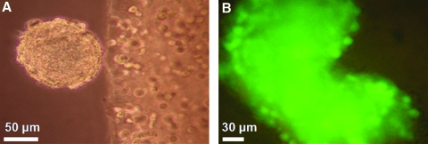
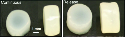
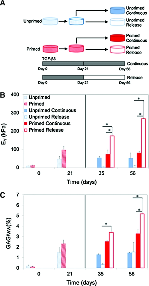
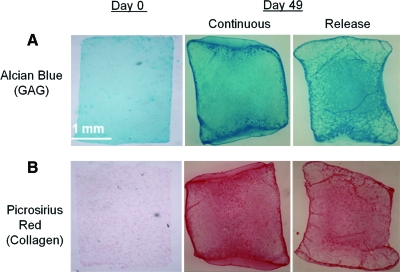
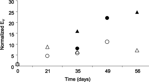
This study investigated the potential use of synovium-derived stem cells (SDSCs) as a cell source for cartilage tissue engineering. Harvested SDSCs from juvenile bovine synovium were expanded in culture in the presence (primed) or absence (unprimed) of growth factors (1 ng/mL transforming growth factor-β(1), 10 ng/mL platelet-derived growth factor-ββ, and 5 ng/mL basic fibroblast growth factor-2) and subsequently seeded into clinically relevant agarose hydrogel scaffolds. Constructs seeded with growth factor-primed SDSCs that received an additional transient application of transforming growth factor-β(3) for the first 21 days (release) exhibited significantly better mechanical and biochemical properties compared to constructs that received sustained growth factor stimulation over the entire culture period (continuous). In particular, the release group exhibited a Young's modulus (267±96 kPa) approaching native immature bovine cartilage levels, with corresponding glycosaminoglycan content (5.19±1.45%ww) similar to native values, within 7 weeks of culture. These findings suggest that SDSCs may serve as a cell source for cartilage tissue engineering applications.


Figures

FIG. 1.
FIG. 1.
Schematic of the study design. Synovium-derived stem cells (SDSCs) were expanded in two-dimensional (2D) culture with (primed) or without (unprimed) growth factors. Once in 3D culture, the culture medium was supplemented with transforming growth factor (TGF)-β3 for the first 21 days (release) or throughout the study (continuous).
FIG. 2.
FIG. 2.
(A) Representative image of aggregated SDSCs emanating from the construct on the right half of the image (20× magnification). (B) Viability of the floating cell mass in the culture media as indicated by pervasive green fluorescence of the cells (40× magnification). Color images available online at www.liebertonline.com/tea
FIG. 3.
FIG. 3.
Representative gross morphology of constructs seeded with growth factor primed synovium cells at day 49 (Study B). The continuous group (left panel) had final average dimensions of φ=4.67±0.23 mm, thickness=2.91±0.11 mm, and the release group (right panel) had final dimensions of φ=4.81±0.19 mm, thickness=2.98±0.11 mm (n=5). Color images available online at www.liebertonline.com/tea
FIG. 4.
FIG. 4.
(A) Schematic of the experimental groups, (B) equilibrium Young's modulus, and (C) glycosaminoglycan (GAG) (%ww) for constructs cultured over 8-week study period. Solid line indicates release at day 21 time point (*p<0.05). Color images available online at www.liebertonline.com/tea
FIG. 5.
FIG. 5.
Representative GAG and collagen staining at days 0 and 49 (A) alcian blue staining (GAG) and (B) picrosirius red staining (collagen) for constructs seeded with growth factor primed SDSCs. Color images available online at www.liebertonline.com/tea
FIG. 6.
FIG. 6.
Representative immunohistochemical staining for (A) type II collagen (green), (B) type VI collagen (green), and (C) negative control image (red for cell nuclei) at day 49 (40× magnification). Color images available online at www.liebertonline.com/tea
FIG. 7.
FIG. 7.
Comparison of the Young's modulus (EY) normalized to day 0 values for the initial study (triangles) and the repeat study (circles). TGF-β3 was added to the culture media either for a transient period of time (release: filled symbols) or continuously throughout the 3D culture (continuous: open symbols). All groups were primed in 2D culture with a growth factor cocktail.

Similar articles

Cited by

References

    1. Guilak F. Sah R.L. Setton L.A. Physical regulation of cartilage metabolism. In: Mow V.C., editor; Hayes W.C., editor. Basic Orthopaedic Biomechanics. New York: Raven Press; 1997. pp. 197–207.
    1. Mow V.C. Bachrach N.M. Setton L.A. Guilak F. Stress, strain, pressure, and flow fields in articular cartilage and chondrocytes. In: Mow V.C., editor; Guilak F., editor; Hayes W.C., editor. Cell Mechanics and Cellular Engineering. New York: Springer-Verlag; 1994. pp. 345–379.
    1. Ronziere M.C. Perrier E. Mallein-Gerin F. Freyria A.M. Chondrogenic potential of bone marrow- and adipose tissue-derived adult human mesenchymal stem cells. Biomed Mater Eng. 2010;20:145. - PubMed
    1. Moutos F.T. Estes B.T. Guilak F. Multifunctional hybrid three-dimensionally woven scaffolds for cartilage tissue engineering. Macromol Biosci. 2010;10:1355. - PMC - PubMed
    1. Lima E.G. Bian L. Ng K.W. Mauck R.L. Byers B.A. Tuan R.S., et al. The beneficial effect of delayed compressive loading on tissue-engineered cartilage constructs cultured with TGF-beta3. Osteoarthritis Cartilage. 2007;15:1025. - PMC - PubMed

Publication types

MeSH terms

Substances