. 2011 May 4:6:22.
doi: 10.1186/1749-8104-6-22.

Rostral growth of commissural axons requires the cell adhesion molecule MDGA2

Affiliations

Rostral growth of commissural axons requires the cell adhesion molecule MDGA2

Pascal Joset et al. Neural Dev. .

Abstract

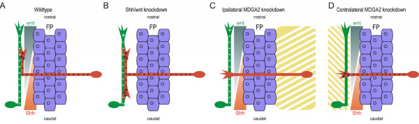
Background: Long-distance axonal growth relies on the precise interplay of guidance cues and cell adhesion molecules. While guidance cues provide positional and directional information for the advancing growth cone, cell adhesion molecules are essential in enabling axonal advancement. Such a dependence on adhesion as well as guidance molecules can be well observed in dorsal commissural interneurons, which follow a highly stereotypical growth and guidance pattern. The mechanisms and molecules involved in the attraction and outgrowth towards the ventral midline, the axon crossing towards the contralateral side, the rostral turning after midline crossing as well as the guidance along the longitudinal axis have been intensely studied. However, little is known about molecules that provide the basis for commissural axon growth along the anterior-posterior axis.

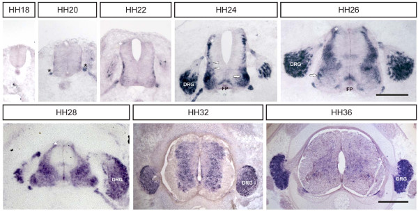
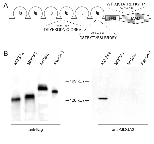
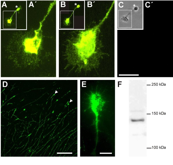
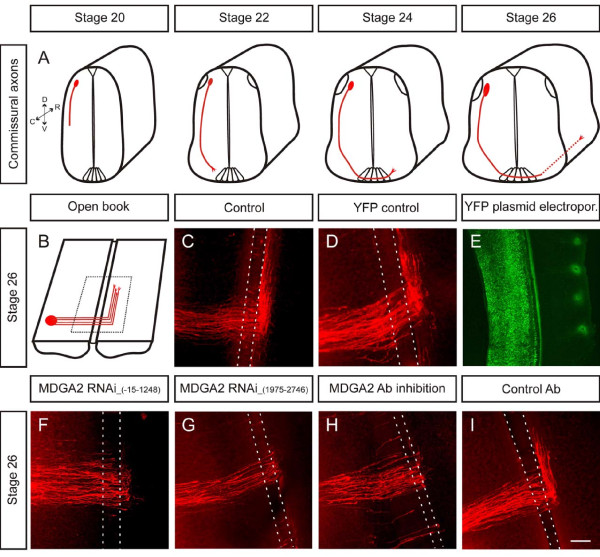
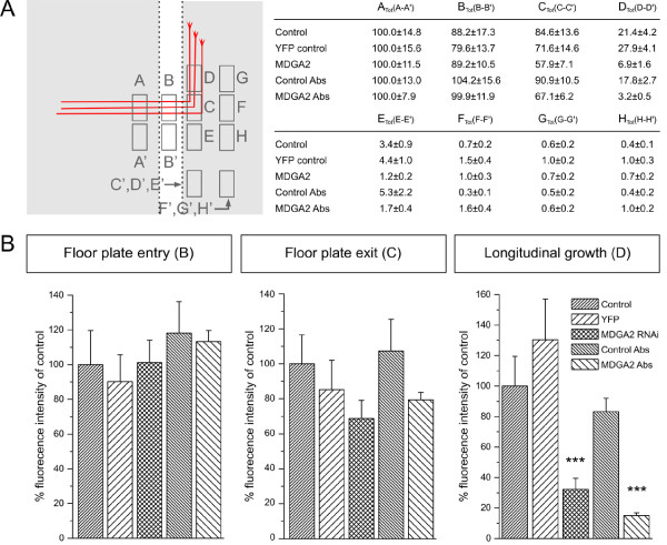
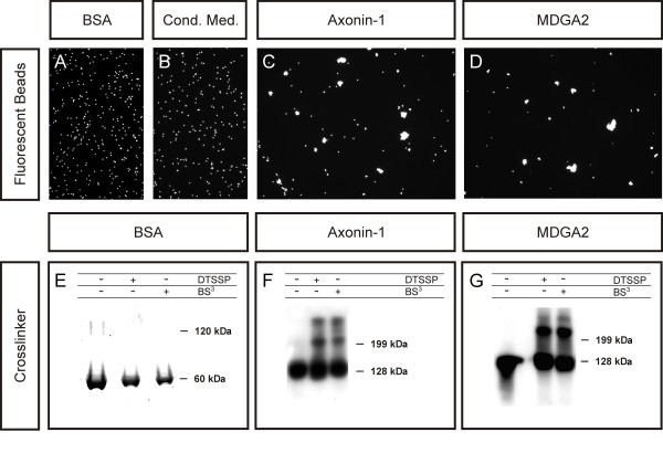
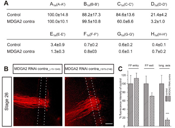
Results: MDGA2, a recently discovered cell adhesion molecule of the IgCAM superfamily, is highly expressed in dorsolaterally located (dI1) spinal interneurons. Functional studies inactivating MDGA2 by RNA interference (RNAi) or function-blocking antibodies demonstrate that either treatment results in a lack of commissural axon growth along the longitudinal axis. Moreover, results from RNAi experiments targeting the contralateral side together with binding studies suggest that homophilic MDGA2 interactions between ipsilaterally projecting axons and post-crossing commissural axons may be the basis of axonal growth along the longitudinal axis.

Conclusions: Directed axonal growth of dorsal commissural interneurons requires an elaborate mixture of instructive (guidance) and permissive (outgrowth supporting) molecules. While Wnt and Sonic hedgehog (Shh) signalling pathways have been shown to specify the growth direction of post-crossing commissural axons, our study now provides evidence that homophilic MDGA2 interactions are essential for axonal extension along the longitudinal axis. Interestingly, so far each part of the complex axonal trajectory of commissural axons uses its own set of guidance and growth-promoting molecules, possibly explaining why such a high number of molecules influencing the growth pattern of commissural interneurons has been identified.


Figures

Figure 1
Figure 1
Expression of MDGA2 mRNA in the chicken spinal cord. Cross-sections of lumbosacral chicken spinal cords were incubated with Dig-labelled MDGA2 antisense RNA and the resulting RNA complexes were visualized using AP-conjugated anti-Dig antibodies. At stage 20, expression is mainly restricted to boundary cap cells at the ventral motor exit points (black asterisks). At stages 24 and 26, MDGA2 mRNA is clearly detectable in commissural interneurons (white asterisks). Starting at stage 24, transcripts can also be observed in dI6/V0 (white arrowheads) and ventrally located V1 interneurons (white arrow) and in a subpopulation of DRG neurons. Between stages 24 and 26 a weak transient expression of MDGA2 transcripts can also be seen in floor-plate cells (FP) and in various interneuron subpopulations. By stage 24, expression of MDGA2 transcripts become up-regulated in different motoneuron subpopulations. Expression levels of MDGA2 transcripts in the spinal cord gradually decrease after stage 28, and by stage 36 only expression in DRG neurons and a subpool of motoneurons persists. The embryonic (Hamburger-Hamilton (HH) stages are indicated. Scale bars represent 200 μm for stages 18 to 26 and 400 μm for stages 28, 32 and 36.
Figure 2
Figure 2
Peptide antibodies specifically recognize the MDGA2 protein. (A) Structural representation of the MDGA2 protein, with the different domains and amino acids chosen for immunization indicated. Immunoglobulin (Ig) domains are represented as open circles, the fibronectin type III repeat (FN3) is depicted as a box and the MAM domain is shown as a hexagonal structure. Two of the peptides used for immunization are located within Ig domains, the third peptide was generated against an amino acid sequence deriving from the MAM domain. (B) Demonstration of MDGA2 antibody specificity. Recombinant flag-tagged MDGA2, MDGA1, NrCAM and axonin-1 were separated by SDS-PAGE and the different proteins were detected by western blotting using either a monoclonal antibody against the flag tag or the newly generated MDGA2 peptide antibodies. While the flag antibody clearly recognizes all recombinant IgCAMs, the pooled MDGA2 peptide antibodies specifically recognize the MDGA2 protein. Note that even when loading higher protein concentrations or using much longer exposure times, no cross-reactivity of the MDGA2 peptide antibodies with other IgCAMs could be detected.
Figure 3
Figure 3
MDGA2 is expressed on the surface of commissural interneurons and dorsal root ganglia neurons. (A-C) Immunolabelling of dissociated dorsal commissural axons. Cultured dorsal commissural neurons express MDGA2 (A,A') as well as the dorsal commissural marker axonin-1 (B,B') on their surface. The position of the cell body is indicated by the asterisk. The growth cone as well as the axonal shaft (A',B') are strongly labelled in both cases whereas a control staining with the secondary antibody only (C') did not yield any labelling. (A',B') Higher magnification images of the axon shaft and growth cone of the neuron seen in (A,B), whereas (C) shows a phase contrast image of the neuron stained with the secondary antibody only (C'). (D,E) Immunolabelling of sensory neurons in culture. While immunostaining using MDGA2 peptide antibodies results in bright labelling of sensory growth cones (D, arrowheads), neuronal shafts are less intensely labelled. (E) Higher magnification image of a sensory growth cone. Note that not only the central part of the growth cone is stained with MDGA2 antibodies but also the extending filopodia. Scale bars: 10 μm in (A-C,E); 100 μm in (D). (F) Western blot analysis of DRG extracts using MDGA2 peptide antibodies. A band of around 135 kDa can be seen in protein extracts from stage 30 DRGs separated by SDS-PAGE and visualized using MDGA2 peptide antibodies.
Figure 4
Figure 4
Down-regulation of MDGA2 causes pathfinding errors of commissural axons. (A) Schematic diagram of the time course of commissural axon pathfinding. Note that midline crossing occurs between stages 22 and 24, and rostral axon turning is initiated around stage 24. (B) Schematic drawing of a stage 26 open-book preparation. The red circle represents the DiI injection site labelling cell bodies of dorsolateral commissural neurons and their axons. (C,D) Confocal images of DiI-injected open-book preparations of an untreated control embryo (C) and an embryo injected and electroporated with a YFP control plasmid (D). Note that in both cases commissural axons make a sharp turn upon leaving the midline area and grow rostrally along the longitudinal axis of the spinal cord. The area between the dashed lines indicates the location of the floor-plate. (E) Electroporation efficiency with YFP. An embryo electroporated with a YFP-plasmid was imaged with epifluorescence. (F,G) RNAi knockdown experiments in which embryos were co-injected with long dsRNA and a YFP expression plasmid. Two independent, non-overlapping fragments were used to produce long dsRNA. Numbers in parentheses indicate the cDNA sequences used to produce dsRNA. Ipsilateral electroporation resulted in a slightly reduced number of axons reaching the contralateral side and a lack of growth of commissural axons along the longitudinal axis. Identical defects were seen with both dsRNA fragments. (H,I) A similar phenotype was observed when MDGA2 peptide antibodies (Ab) were injected into the central canal of stage 20 chicken embryos (H), whereas normal axon growth was seen in embryos injected with a control IgG (I). Scale bars: 100 μm.
Figure 5
Figure 5
Quantification of commissural axon outgrowth defects after perturbation of MDGA2 function by RNAi and antibody injection. (A) Schematic representation of the areas used to measure fluorescence intensities in control, RNAi and antibody (Ab)-treated embryos. A, ipsilateral; B, floor plate; C, floor-plate exit; D, rostral longitudinal axis; E, caudal turn; F, no turn; G, 45° turn; H, -45° turn. Background fluorescence (A', B', C'...) at corresponding locations were subtracted from the values obtained at the different measurement sites (A, B, C....). The table shows the normalized intensities, with Atot (A - A') of the control situation set as the reference value (100; for details see Materials and methods). Note that the fluorescent intensities (at sites B, C, D...) for all experimental conditions are adjusted by the same factor as calculated by the normalization at the site Atot. (B) Data obtained by measurements of fluorescence intensity are presented as histograms to indicate the percentages of fluorescence intensity at particular locations. Values for control embryos were set to 100% at each of the analyzed sites. While in all analyzed conditions commissural axons were able to enter the floor plate (B, left panel), in MDGA2 RNAi-treated, as well as in embryos injected with the MDGA2 antibody fewer axons exited at the contralateral site (B, middle panel). The strongest difference between control and experimental animals was seen in commissural axon growth along the longitudinal axis, with only 15 to 30% of the axons extending rostrally after perturbation of MDGA2 function (B, right panel). While the effect on commissural axon stalling in the floor plate was not statistically significant, the lack of commissural axon growth along the longitudinal axis was highly significant (***P < 0.005; standard t-test). Error bars are given as standard errors (SEM).
Figure 6
Figure 6
MDGA2 is capable of interacting homophilically. Two independent assays were used to analyze the binding capabilities of MDGA2. In an aggregation assay, fluorescent beads were coupled with BSA, proteins from conditioned medium of non-transfected cells, recombinant axonin-1 or recombinant MDGA2 and analyzed for their aggregation behaviour. (A-D) While no aggregates were observed with BSA (A) or beads coated with proteins released by mock-transfected cells (B), strong aggregation was detected with axonin-1- (C) and MDGA2-coupled beads (D). (E-G) Similar results were seen when chemical cross-linkers were used to monitor the binding capabilities of these molecules. (E) Silver staining of gels indicated that, in a concentrated BSA solution, no high molecular weight aggregates were formed in the presence of the chemical cross-linkers. Crosslinking of conditioned media containing recombinant flag-tagged axonin-1 (F) or flag-tagged MDGA2 (G) resulted in a molecular weight shift of a substantial part of the detected protein as observed by western blot assays using anti-flag antibodies. In the case of axonin-1, note that two additional bands are observed in the presence of chemical cross-linkers, indicating that axonin-1 is capable of forming multimeric complexes.
Figure 7
Figure 7
Ipsilaterally and contralaterally projecting tracts co-localise in the ventral funiculus. (A,B) The lipophilic dyes DiI (red) and DiA (green) were injected into contra- and ipsilaterally projecting neurons, respectively. Subsequently, the tissue was cut into 25-μm thick sections. Panel (B) represents the plane of dye injection, and the section shown in (C-E) was 100 μm more rostral relative to (B) (schematic drawing in (A) labels the sections). (C) Post-crossing axons of DiI-labelled dorsal interneurons project in the contralateral ventral funiculus (arrowhead). (D) DiA-labelled ipsilaterally projecting axons are found throughout the ventral funiculus, including the medial part close to the floor plate, where dorsal interneurons turn into the longitudinal axis (arrowhead). (E) An overlay of contra- and ipsilaterally projecting axons shows that these populations co-localize in the ventral funiculus (arrowhead). Note that due to the juxtaposition of the dorsal interneurons and the dorsal funiculus, injection of DiI also stained the longitudinal axonal tracts of the dorsal funiculus (asterisk in (B,C,E)). Ca, caudal; Ro, rostral. Scale bar: 200 μm.
Figure 8
Figure 8
Down-regulation of MDGA2 on the contralateral side causes phenotypes similar to those seen in ipsilateral knockdowns. Embryos were injected with either of the two non-overlapping long dsRNA fragments covering the sequences -15 to 1,248 and 1,975 to 2,746, respectively. Following contralateral electroporation, embryos treated with either fragment did show severe defects in commissural axon growth. (A,C) While the table (A) gives the fluorescent intensities measured at different locations in control and RNAi-treated embryos (for details see Figure 4 and Materials and methods), the histogram (C) depicts the normalized fluorescent intensities in percentage of the control. In analogy to the results seen with ipsilateral electroporations of MDGA2 dsRNA (Figure 4), rostral turning of commissural axons after midline crossing in contralateral RNAi knockdown embryos is strongly reduced compared to control embryos. (B) Representative confocal pictures of open-book preparations of embryos after MDGA2 knockdown are shown. Most post-crossing commissural axons stalled at the floor-plate exit site and did not grow along the longitudinal axis. FP, floor-plate. Error bars given as standard errors (SEM). Scale bar: 100 μm.
Figure 9
Figure 9
Mechanism of commissural axon growth and guidance after midline crossing. Schematic drawings of the commissural axon trajectory under various experimental conditions. Dorsally located commissural interneurons are depicted in red, whereas ipsilaterally projecting interneurons are shown in green. Wnt (dark green) and Shh (light red) gradients are indicated. Expression sites for MDGA2 are highlighted by the plus signs. Areas of MDGA2 knockdown are indicated by the yellow striped regions. (A) Under control conditions commissural axons turn rostrally to grow along the longitudinal axis towards the brain. (B) In embryos lacking rostrocaudal activity gradients of the guidance cues Wnt4 or Shh, commissural axons either stall or turn randomly either rostrally or caudally. (C,D) Ipsilateral knockdown of MDGA2 prevents commissural axonal growth along the longitudinal axis (C), a phenotype also seen after contralateral knockdown of MDGA2 (D). FP, floor-plate.

Similar articles

Cited by

References

    1. Chilton JK. Molecular mechanisms of axon guidance. Dev Biol. 2006;292:13–24. doi: 10.1016/j.ydbio.2005.12.048. - DOI - PubMed
    1. Kaprielian Z, Runko E, Imondi R. Axon guidance at the midline choice point. Dev Dyn. 2001;221:154–181. doi: 10.1002/dvdy.1143. - DOI - PubMed
    1. Bentley D, Caudy M. Pioneer axons lose directed growth after selective killing of guidepost cells. Nature. 1983;304:62–65. doi: 10.1038/304062a0. - DOI - PubMed
    1. Palka J, Whitlock KE, Murray MA. Guidepost cells. Curr Opin Neurobiol. 1992;2:48–54. doi: 10.1016/0959-4388(92)90161-D. - DOI - PubMed
    1. Stoeckli ET. Molecular mechanisms of commissural axon pathfinding. Prog Brain Res. 1998;117:105–114. - PubMed

Publication types

MeSH terms

LinkOut - more resources