. 2011 Jun 10;10(6):586-94.
doi: 10.1016/j.dnarep.2011.03.006. Epub 2011 May 4.

The budding yeast Mei5-Sae3 complex interacts with Rad51 and preferentially binds a DNA fork structure

Affiliations

The budding yeast Mei5-Sae3 complex interacts with Rad51 and preferentially binds a DNA fork structure

Amanda F Say et al. DNA Repair (Amst). .

Abstract

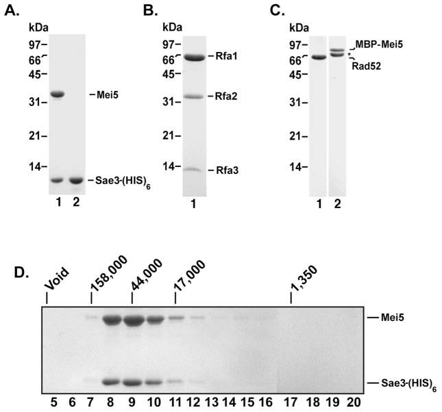
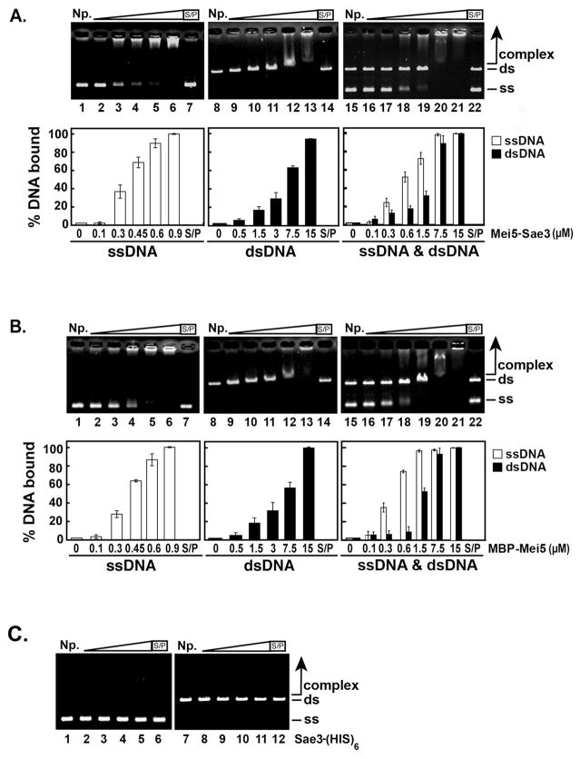
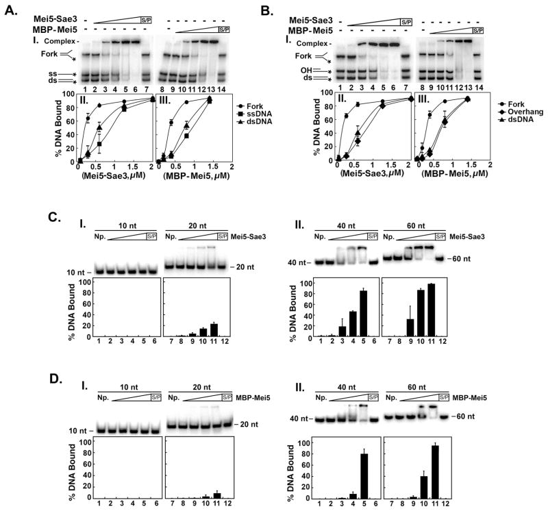
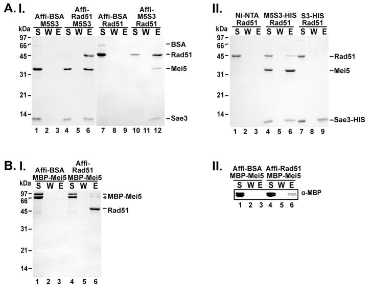
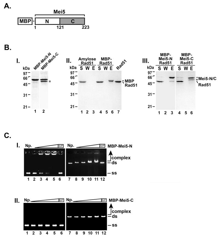
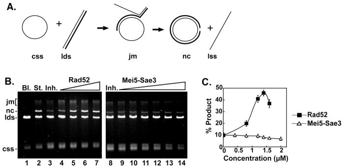
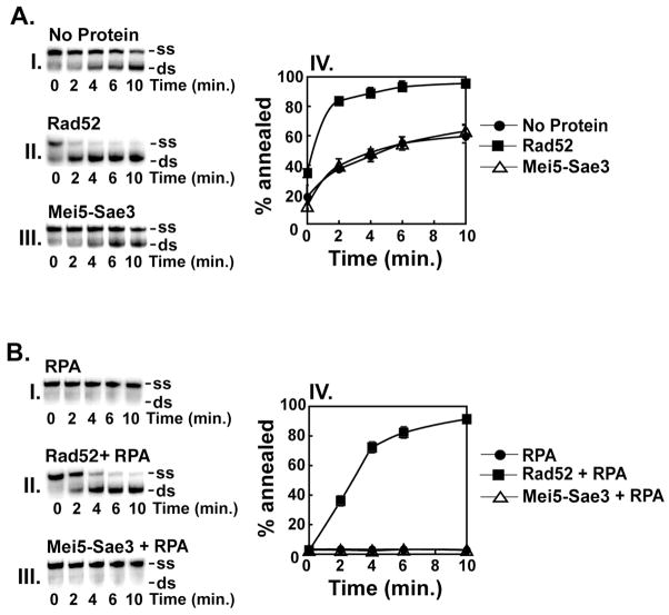
Meiotic homologous recombination in Saccharomyces cerevisiae involves formation of nucleoprotein filaments of Rad51 and Dmc1 that mediate DNA strand exchange between homologous chromosomes. The Mei5-Sae3 protein complex functions as a recombination mediator to promote nucleation of the Dmc1 recombinase onto replication protein A-coated single-stranded DNA. Here, we have expressed and purified the Mei5 protein, Sae3 protein and the Mei5-Sae3 complex for biochemical studies. We show the Mei5-Sae3 complex preferentially binds a fork-like DNA substrate to 3' overhanging DNA, single-stranded DNA or double-stranded DNA. We demonstrate that Mei5 confers DNA binding activity to the Mei5-Sae3 complex. We determined Mei5-Sae3 interacts with the Rad51 recombinase through the N-terminal domain of Mei5. Unlike Rad52, Mei5-Sae3 lacks recombination mediator activity for Rad51. Importantly, we find that the Mei5-Sae3 complex does not harbor single-strand DNA annealing activity. These properties of the Mei5-Sae3 complex distinguishes it from the Rad52 protein, which serves as the mediator of Rad51 and is involved in the single-strand DNA annealing pathway of homologous recombination.


Conflict of interest statement

Conflicts of interest

The authors declare that there are no conflicts of interest.

Figures

Figure 1
Figure 1. Purified Mei5-Sae3, Sae3, RPA, Rad52, and MBP-Mei5 proteins and stoichiometry of the Mei5-Sae3 complex
Mei5-Sae3 complex (1.5 μg; A, lane 1), Sae3-(HIS)6 (1.5 μg; A, lane 2), RPA (3 μg; B), Rad52 (1 μg; C, lane 1) and MBP-Mei5 (1 μg; C, lane 2) were resolved by 12% SDS-PAGE polyacrylamide gel and stained with Coomassie Blue. (*) Indicates the C-terminal truncation product of MBP-Mei5. The Mei5-Sae3 complex (D) was filtered through a 20 mL Sephacryl S-100 column. Fractions were TCA precipitated and subjected to SDS-PAGE on 12% polyacrylamide gels and stained with Coomassie Blue. Protein standards (Bio-Rad) bovine thyroglobulin (670 kDa, Void), bovine γ-globulin (158 kDa), chicken ovalbumin (44 kDa), horse myoglobin (17 kDa) and vitamin B12 (1.35 kDa) were filtered on a 20 mL Sephacryl S-100 column and eluted in the indicated fractions.
Figure 2
Figure 2. DNA binding activity of Mei5-Sae3, Mei5 and Sae3 with φX174 DNA
The indicated concentrations of Mei5-Sae3 (A) or MBP-Mei5 (B) were incubated with φX174 ssDNA (30 μM nucleotides, lanes 2–7), linearized φX174 RF dsDNA (30 μM base pairs, lanes 9–14), or both ssDNA and dsDNA (30 μM nucleotides and 30 μM base pairs, respectively, lanes 16–22) at 37°C for 10 min. (C) Sae3-(HIS)6 protein (1 μM, lanes 2 and 8; 5 μM, lanes 3 and 9; 15 μM, lanes 4 and 10; 30 μM, lanes 5, 6, 11 and 12) was incubated with φX174 ssDNA (30 μM nucleotides, lanes 2–6) or linearized φX174 RF dsDNA (30 μM base pairs, lanes 8–12) at 37°C for 10 min. The reaction products were separated on 0.9 % agarose gels and stained with ethidium bromide. The gels were quantified using Quantity One (Bio-Rad) software. Where indicated, the reaction was treated with SDS (0.5% final) and Proteinase K (0.5 mg/mL) at 37°C for 15 min prior to analysis. S/P, SDS/Proteinase K; NP, no protein control; ss, ssDNA; ds, dsDNA. The average values from 4 experiments were plotted (A and B, bottom panels).
Figure 3
Figure 3. DNA binding specificity of Mei5-Sae3 and Mei5
(A and B) Mei5-Sae3 (panel I; 0.1 μM, lane 2; 0.3 μM, lane 3; 0.6 μM, lane 4; 1.25 μM, lane 5; 2.0 μM, lane 6 and 7) and MBP-Mei5 (panel I; 0.07 μM, lane 9; 0.2 μM, lane 10; 0.4 μM, lane 11; 0.83 μM, lane 12; 1.38 μM, lane 13 and 14) were incubated with 0.05 pmol of radiolabeled fork, dsDNA and ssDNA (A, panel I) or radiolabeled fork, dsDNA, and OH (B, panel I) for 10 min at 37°C and subjected to non-denaturing-PAGE. The gels were dried and visualized using a Typhoon phosphorimager. Reactions with Mei5-Sae3 (A and B, panels II) and MBP-Mei5 (A and B, panel III) were quantified using ImageQuant (GE Healthcare) software. (C and D) Mei5-Sae3 and MBP-Mei5, respectively (panel I and II; 0.08 μM, lane 2 and 8; 0.26 μM, lane 3 and 9; 0.52 μM, lane 4 and 10; 1.3 μM, lane 5 and 11; 1.3 μM, lane 6 and 12) were incubated with 0.05 pmol of radiolabeled poly dT 10-mer (panel I, lanes 1–6), poly dT 20-mer (panel I, lanes 7–12), poly dT 40-mer (panel II, lanes 1–6), and poly dT 60-mer (panel II, lanes 7–12) for 10 min at 37°C and treated as described above. Where indicated, the reaction was treated with SDS (0.5% final) and Proteinase K (0.5 mg/mL) at 37°C for 10 min prior to analysis. S/P, SDS/Proteinase K; NP, no protein control; ss, ssDNA; ds, dsDNA.
Figure 4
Figure 4. Mei5-Sae3 interacts with Rad51 through Mei5
(A) Mei5-Sae3 (panel I) was mixed with Affi-Gel containing covalently conjugated BSA or Rad51 (lanes 1–6). Rad51 was mixed with Affi-Gel containing covalently conjugated BSA or Mei5-Sae3 (lanes 7–12). After a wash, the bound protein was eluted with SDS. The supernatant (S), wash (W), and eluate (E) were separated on a SDS-PAGE gel and stained with Coomassie Blue. (A, panel II) Mei5-Sae3 or Sae3-(HIS)6 (panel II) was incubated separately with Rad51. Nickel-NTA beads were added to the reactions and with Rad51 alone with agitation to capture protein complexes. After a wash, the bound protein was eluted and samples were analyzed as described above. (B) MBP-Mei5 (panel I, lanes 1–6) was mixed with Affi-Gel containing covalently conjugated BSA (panel I lanes 1–3) or Rad51 (panel I lanes 4–6). After a wash, the bound protein was eluted and samples were analyzed as described above. Western analysis using anti–MBP antibodies was used to confirm the presence of MBP-Mei5 protein (panel II, lanes 1–6).
Figure 5
Figure 5. The N-terminal domain of Mei5 interacts with Rad51 and binds DNA
(A) Domain schematic of Mei5. (B) MBP-Mei5-N (1.3 μg; panel I, lane 1) and MBP-Mei5-C (1.2 μg; panel I, lane 2) were resolved using a 12% SDS-PAGE polyacrylamide gel and stained with Coomassie Blue. Rad51 (7 μg) was incubated with MBP (6.5 μg; panel II, lanes 4–6), MBP-Mei5-N (7.0 μg; panel III, lanes 1–3), MBP-Mei5-C (7.0 μg; panel III, lanes 4–6) for 30 min at 4°C. Amylose beads were added to the reactions and with Rad51 alone (panel II, lanes 1–3) for 30 min at 4°C with agitation to capture protein complexes. The supernatant (S) that contained unbound proteins, wash (W), and SDS eluate (E) were analyzed by SDS-PAGE and stained with Coomassie Blue. (C) MBP-Mei5-N and MBP-Mei5-C (panel I and panel II, respectively; 0.55 μM, lanes 2 and 8; 1.66 μM, lanes 3 and 9; 2.77 μM, lanes 4 and 10; 5.5 μM, lanes 5, 6, 11 and 12) were incubated with φX174 ssDNA (30 μM nucleotides, lanes 2–6) and linearized φX174 RF dsDNA (30 μM base pairs, lanes 7–12) at 37°C for 10 min. The reaction products were separated on 0.9 % agarose gels and stained with ethidium bromide. Where indicated, the reaction was treated with SDS (0.5% final) and Proteinase K (0.5 mg/mL) at 37°C for 15 min prior to analysis. S/P, SDS/Proteinase K; NP, no protein control; ss, ssDNA; ds, dsDNA. (*) Indicates the C-terminal truncation product of MBP-Mei5.
Figure 6
Figure 6. Mei5-Sae3 does not mediate Rad51 strand exchange
(A) Schematic representation of the 3-strand homologous DNA pairing and strand exchange reaction. Homologous pairing between the circular ssDNA (css) and linear duplex DNA (lds) substrates yields a DNA joint molecule (jm), which is converted into a nicked circular duplex molecule (nc) by DNA strand exchange liberating linear ssDNA (lss). (B) The standard reaction (St., lane 2) resulted from pre-incubating the ssDNA with Rad51 (9.3 μM) to allow for the formation of presynaptic filaments before the addition of RPA (1.7 μM). Co-incubation of the ssDNA with Rad51 (9.3 μM) and RPA (1.7 μM) resulted in greatly inhibited DNA strand exchange (Inh., lane 3 and 8). Rad52 (0.8 μM, lane 4; 1.2 μM, lane 5; 1.4 μM, lane 6; 1.6 μM, lane 7) or Mei5-Sae3 (0.4 μM, lane 9, 0.8 μM, lane 10; 1.2 μM, lane 11; 1.6 μM, lane 12; 2.0 μM, lane 13) was included during the incubation of ssDNA with Rad51 and RPA. Bl. indicates no protein was added. (C), the results of (B) were plotted.
Figure 7
Figure 7. Mei5-Sae3 lacks DNA single-strand annealing activity
(A) The radiolabeled OL83 and unlabeled OL83-c oligonucleotides (0.83 μM nucleotides) were incubated in the absence of protein (A, panel I) or in the presence of Rad52 (0.34 μM, A, panel II) or Mei5-Sae3 (0.34 μM, A, panel III) at 37°C for the indicated times. (B) The same oligonucleotide substrates (0.83 μM nucleotides) were incubated in the presence of RPA (0.12 μM, B, panel I) and either Rad52 (0.34 μM, B, panel II) or Mei5-Sae3 (0.34 μM, B, panel III) at 37°C for the indicated times. For both (A) and (B), the samples were quenched with excess OL83-c and deproteinized by treatment with SDS (0.5% final), Proteinase K (0.5 mg/mL) for 15 min at 37°C. The reaction mixtures were resolved in 12 % non-denaturing TAE polyacrylamide gels. The gels were dried and subjected to phosphorimaging analysis. The position of the ssDNA (ss) and annealed dsDNA (ds) product are indicated on the right in (panels I–III). The results in panels I–III were quantified with ImageQuant (GE Healthcare) and plotted (panel IV).

References

    1. Keeney S, Giroux CN, Kleckner N. Meiosis-specific DNA double-strand breaks are catalyzed by Spo11, a member of a widely conserved protein family. Cell. 1997;88:375–384. - PubMed
    1. Shinohara A, Ogawa H, Ogawa T. Rad51 protein involved in repair and recombination in S. cerevisiae is a RecA-like protein. Cell. 1992;69:457–470. - PubMed
    1. Bishop DK, Park D, Xu L, Kleckner N. DMC1: a meiosis-specific yeast homolog of E. coli recA required for recombination, synaptonemal complex formation, and cell cycle progression. Cell. 1992;69:439–456. - PubMed
    1. San Filippo J, Sung P, Klein H. Mechanism of eukaryotic homologous recombination. Annu Rev Biochem. 2008;77:229–257. - PubMed
    1. Sung P, Klein H. Mechanism of homologous recombination: mediators and helicases take on regulatory functions. Nat Rev Mol Cell Biol. 2006;7:739–750. - PubMed

Publication types

MeSH terms