. 2011 Jul 22;286(29):25963-72.
doi: 10.1074/jbc.M110.217414. Epub 2011 May 3.

Structural and biochemical characterization of Staphylococcus aureus clumping factor B/ligand interactions

Affiliations

Structural and biochemical characterization of Staphylococcus aureus clumping factor B/ligand interactions

Vannakambadi K Ganesh et al. J Biol Chem. .

Abstract

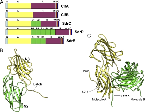
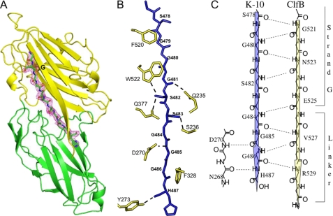
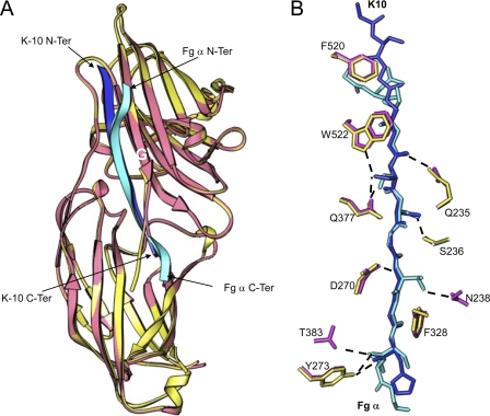
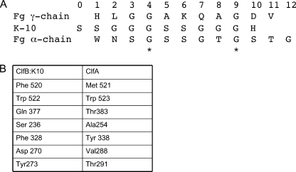
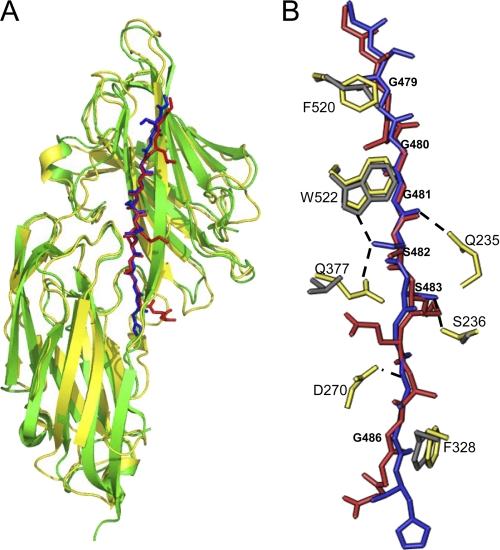
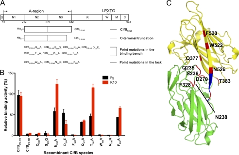
Clumping factor B (ClfB) from Staphylococcus aureus is a bifunctional protein that binds to human cytokeratin 10 (K10) and fibrinogen (Fg). ClfB has been implicated in S. aureus colonization of nasal epithelium and is therefore a key virulence factor. People colonized with S. aureus are at an increased risk for invasive staphylococcal disease. In this study, we have determined the crystal structures of the ligand-binding region of ClfB in an apo-form and in complex with human K10 and Fg α-chain-derived peptides, respectively. We have determined the structures of MSCRAMM binding to two ligands with different sequences in the same site showing the versatile nature of the ligand recognition mode of microbial surface components recognizing adhesive matrix molecules. Both ligands bind ClfB by parallel β-sheet complementation as observed for the clumping factor A·γ-chain peptide complex. The β-sheet complementation is shorter in the ClfB·Fg α-chain peptide complex. The structures show that several residues in ClfB are important for binding to both ligands, whereas others only make contact with one of the ligands. A common motif GSSGXG found in both ligands is part of the ClfB-binding site. This motif is found in many human proteins thus raising the possibility that ClfB recognizes additional ligands.


Figures

FIGURE 1.
FIGURE 1.
Representation of apoClfBN2N3(199–542). A, domain organization of Clf-Sdr proteins. S, signal peptide; A, ligand binding region; B, homologous repeats; R, SD repeats; W, wall-spanning region; M, membrane anchor; C, cytoplasmic tail. B, ribbon representation of structure of the apo-form of ClfB. The N2 and N3 subdomains are shown in green and yellow, respectively. C, representation of the two symmetry-related molecules in the unit cell. The latch of the molecule A (yellow) is inserted in the latching trench of molecule B (green) and vice versa.
FIGURE 2.
FIGURE 2.
Crystal structure of the ClfBN2N3·K10 peptide complex. A, ribbon representation of the ClfB·K10 peptide complex. The peptide is shown in stick format and is colored by atom type: carbon, green; nitrogen, blue; and oxygen, red. 2FoFc electron density map around the peptide contoured at 1σ is shown in purple. B, representation of the key side-chain interactions of ClfB and the K10 peptide. The K10 peptide is shown in blue stick format. Side-chain atoms of interacting residues in ClfB are shown in yellow. Hydrogen bonds are shown as dotted lines. C, schematic representation of the backbone hydrogen bonding interactions between ClfB and the K10 peptide. Hydrogen bonds are shown as dotted lines. The K10 peptide makes a parallel β-sheet complementation with ClfB.
FIGURE 3.
FIGURE 3.
Comparison of ClfB·K10 and ClfB·Fg α-chain peptide. A, ribbon representation of the superimposition of ClfB·K10 and ClfB·Fg α-chain complexes. ClfB from the K10 and Fg α-chain complexes is shown in yellow and pink, respectively. The K10 peptide is shown in blue, and the Fg α-chain is shown in cyan. B, comparison of the key interactions of Fg α-chain and K10 with ClfB. The ClfB molecules of the two complexes were superimposed and are not shown for clarity. K10 peptide is shown in blue and the Fg α-chain peptide is shown in cyan. The side chains of ClfB from the K10 complex are shown in yellow, and those from Fg α-chain complex are shown in pink.
FIGURE 4.
FIGURE 4.
Structure-based alignment of the sequences of ClfB·ligand complexes and the ClfA·Fg γ-chain peptide complex. A, alignment of the amino acid sequences of the ligand molecules based on the structure of their complexes. Each of these amino acid-binding subsites within the binding site were designated 0–12 starting from the N terminus. The ClfB·K10 and ClfB·Fg α-chain peptide complexes and the ClfA·Fg γ-chain peptide complex were superimposed, and the aligned sequences are listed. B, table of the key ligand-binding residues in ClfB and the corresponding residues in ClfA.
FIGURE 5.
FIGURE 5.
Superimposition of ClfB·K10 and ClfA·Fg γ-chain peptide complexes. A, ribbon representation of the superimposition of the ClfB·10 and ClfA·Fg γ-chain-peptide complexes. ClfA and ClfB molecules are shown in green and yellow, respectively. The K10 peptide is shown in blue, and the Fg γ-chain peptide is shown in red. B, comparison of the key side-chain interactions of the ClfB·K10 peptide and ClfA·Fg γ-chain peptide complexes. The K10 peptide is shown as a blue stick object. The Fg γ-chain peptide from the ClfA·γ-chain complex (18) has been overlaid on the K10 peptide and is shown as a red stick object. Side-chain atoms of interacting residues in ClfB are shown in yellow, and the corresponding residues in ClfA are shown in gray. Hydrogen bonds are shown as dotted lines.
FIGURE 6.
FIGURE 6.
Illustration of ClfB domains and recombinant ClfB proteins affinities for K10 and Fg. A, schematic representation of the domain structure of ClfB. S, signal sequence; A-region, containing N1, N2, and N3; R, serine-aspartic acid repeat region; W, wall-spanning fragment; M, transmembrane domain; C, cytoplasmic tail; LPXTG, cell wall anchoring motif. Also shown are the His-tagged truncated proteins used in this study. B, relative binding affinities of different ClfB recombinant protein species for Fg and K10. Fg and recombinant K10 were immobilized on Immulon 4BH plates, and increasing concentrations of ClfB recombinant protein species were added to the wells. Bound protein was detected with rabbit anti-ClfBN2N3 followed by an anti-rabbit HRP-labeled antibody. The binding constants were analyzed by nonlinear regression for one binding site (GraphPad Prism). The relative binding activity of the mutant recombinant ClfB species were calculated as percent of the wild type protein binding. C, positions of the amino acid mutations carried out for characterization of the ligands binding to ClfB are plotted on the Ribbon model of ClfB. The N2 and the N3 subdomains of ClfB are shown in green and blue, respectively. The lock residues are shown in blue, and the positions of the point mutation are shown in red and labeled.

Similar articles

Cited by

References

    1. Diekema D. J., Pfaller M. A., Schmitz F. J., Smayevsky J., Bell J., Jones R. N., Beach M. (2001) Clin. Infect. Dis. 32, S114–S132 - PubMed
    1. Lowy F. D. (1998) N. Engl. J. Med. 339, 520–532 - PubMed
    1. Chambers H. F. (2009) J. Infect. Dis. 199, 291–293 - PubMed
    1. Kluytmans J., van Belkum A., Verbrugh H. (1997) Clin. Microbiol. Rev. 10, 505–520 - PMC - PubMed
    1. von Eiff C., Becker K., Machka K., Stammer H., Peters G. (2001) N. Engl. J. Med. 344, 11–16 - PubMed

Publication types

MeSH terms

LinkOut - more resources